
Figures & data
Table 1. Demographic and clinical characteristics in patients with active tuberculosis (TB) and healthy control individuals
Figure 1. Effect of Mycobacterium tuberculosis (Mtb) infection on human monocytes in pulmonary tuberculosis (TB) patient. Human peripheral blood mononuclear cells (PBMCs) were isolated from healthy control (HC; n = 34) and pulmonary TB patients (TB; n = 34). (a) CD14-positive (CD14+) moncytes were sorted and determined by fluorescence-activated cell sorting (FACS). (b) Apoptosis positive cells in above primary moncytes was measured on FACS. All operations were performed in triplicate and * P < 0.05
Figure 2. Expression of miR-125b-5p in human primary moncytes and macrophages infected by H37Rv in vitro. Relative expression level of miR-125b-5p was measured with real-time PCR in (a) PBMCs from HC and TB patients, and (b, c) H37Rv-infected human peripheral blood monocytes-derived macrophages (MDMs) and THP-1 cells-derived macrophages (TDMs) for 48 h. (d) The mycobacterial viability in transfected MDMs and TDMs after H37Rv infection was determined by colony-forming units (CFU) assay. (e) FACS examined apoptosis rate of H37Rv-infected MDMs and TDMs. (f-h) Enzyme-linked immunosorbent assay (ELISA) measured levels of inflammatory factors, interleukin (IL)-6, IL-10, and tumor necrosis factor (TNF)-α in cell culture supernatant of H37Rv-infected MDMs and TDMs. (i) Western blot measured levels of apoptosis-related proteins, B cell lymphoma (Bcl)-2, Bcl-2-associated X Protein (Bax), and Bcl-2-interacting mediator (Bim) in H37Rv-infected MDMs and TDMs. All experiments were performed in triplicate and * P < 0.05
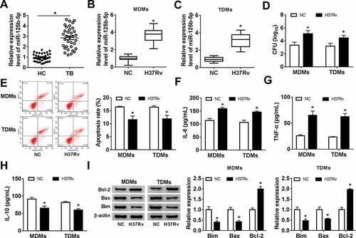
Figure 3. Effect of miR-125b-5p blockage on human macrophage apoptosis in vitro with H37Rv infection. (a) Real-time PCR detected miR-125b-5p expression level in MDMs and TDMs transfected with miR-125b-5p inhibitor (in-miR-125b-5p) or its negative control (in-miR-NC). (bh) Above transfected MDMs and TDMs at 48 h were then subjected with H37Rv infection for 48 h. (b) Real-time PCR detected miR-125b-5p expression level. (c) CFU assay determined mycobacterial viability. (d) Apoptosis rate was recorded by FACS. (e–g) ELISA measured IL-6, IL-10 and TNF-α levels in cell culture supernatant. (h) Western blot assay examined Bcl-2, Bax and Bim levels with normalization to β-actin. All operations were carried out at least three times and * P < 0.05
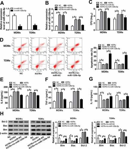
Figure 4. DNA damage-regulated autophagy modulator 2 (DRAM2) was a target gene of miR-125b-5p in human macrophages. (a) The potential binding sequences of hsa-miR-125b-5p in wild type of DRAM2 3ʹ UTR (DRAM2-WT) was presented and mutated to construct the mutation of DRAM2 3ʹ UTR (DRAM2-MUT). (b-e) Dual-luciferase reporter assays assessed luciferase activity of plasmid carrying DRAM2-WT/MUT in MDMs and TDMs co-transfected with (b, c) miR-125-5p/NC mimic (miR-125-5p/NC) or (d, e) in-miR-125-5p/NC inhibitor. (f-h) Real-time PCR measured DRAM2 expression in (f) human primary moncytes from TB and HC, and (g, h) macrophages (MDMs and TDMs) with H37Rv infection or not. (i) Western blot assay examined expression level of DRAM2 in MDMs and TDMs transfected with miR-NC, miR-125-5p, in-miR-NC, or in-miR-125-5p. (j) The correlation analysis was conducted between miR-125b-5p and DRAM2 expression with Spearman rank correlation test. All operations were carried out in triplicate. * P < 0.05
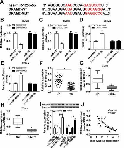
Figure 5. The promotion of miR-125b-5p knockdown on human macrophages apoptosis in vitro with H37Rv infection was abolished by silencing DRAM2. (a) Western blot assay examined expression level of DRAM2 in MDMs and TDMs transfected with siRNA against DRAM2 (si-DRAM2) or its negative control (si-NC). (bg) MDMs and TDMs were transfected with in-miR-125b-5p alone or combined with si-DRAM2 for 48 h, followed with H37Rv infection for 48 h. (b) CFU assay determined mycobacterial viability. (c) Apoptosis rate was recorded by FACS. (d–f) ELISA measured IL-6, IL-10, and TNF-α levels in cell culture supernatant. (g) Western blot assay examined Bcl-2, Bax, and Bim levels. All operations were launched in triplicate. * P < 0.05
Supplemental Material
Download Zip (102.2 KB)Availability of data and materials
All original data and materials are available from the corresponding author upon request.