
Figures & data
Figure 1. The expression level of HMGA2 in HUVEC under hypoxic condition
(a) The expression of hypoxia markers HIF-1α and HO-1 increased under hypoxia stimulation for 12 h(n = 6). (b) Protein expression level of HMGA2 in HUVEC with or without hypoxia treatment was shown in western blotting and was normalized to GAPDH (n = 6). (c) The mRNA expression of HMGA2 was detected by PCR (n = 6). (d) HMGA2 expression in HUVEC was revealed by immunofluorescence staining (n = 6). *P < 0.05 vs. control group. All quantitative data were presented as the mean ± SD.
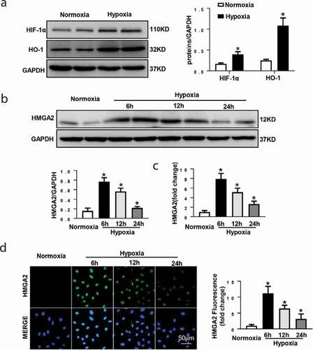
Figure 2. HMGA2 overexpression accelerated hypoxia-induced cell migration and attenuated hypoxia-induced apoptosis in HUVECs
(a) HMGA2 expression of HUVECs in indicated group and the protein expression level was normalized to GAPDH(n = 6). (b)The viability of each group was detected by MTT(repeat 6 times). (c) In the wound healing assay, HUVECs were treated with Ad-GFP or Ad-HMGA2 vector and/or hypoxia for 24 hours and photographed at 0, 12, and 24 hours (n = 6). (d) Representative images (upper panels) and quantification (lower panels) of cell migration under hypoxia 24 h in Transwell chamber assay(n = 6). (e) Caspase3 activity of HUVECs in indicated group with hypoxia treatment for 12 h(n = 6). (f) TUNEL staining exhibited the apoptosis in the HUVECs after infection of Ad-GFP or Ad-HMGA2 vector with or without 12 h hypoxia stimuli (n = 6). (g) Western blotting analyses of the apoptosis associated proteins Bax and Bcl-2 under hypoxia treatment for 12 h(n = 6). *P < 0.05 vs. Ad-GFP+nomoxia group. #P < 0.05 vs. Ad-GFP+hypoxia group. All quantitative data were presented as the mean ± SD.
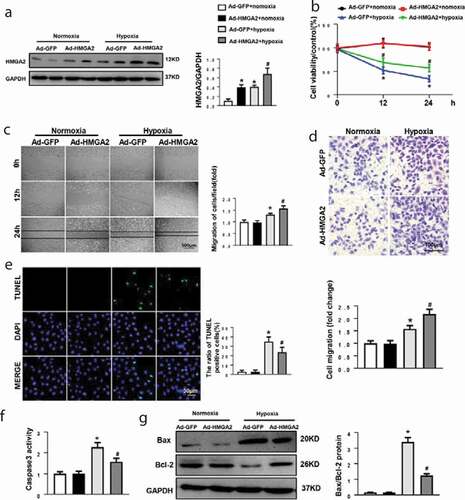
Figure 3. HMGA2 knockdown suppressed hypoxia-induced cell migration and aggravated hypoxia-induced apoptosis in HUVECs
(a) HMGA2 expression in the HUVECs in indicated group and protein expression level was normalized to GAPDH(n = 6). (b) The viability of each group was detected by MTT(repeat 6 times). (c) In the wound healing assay, HUVECs were treated with si-NC or si-HMGA2 and/or hypoxia for 24 hours and photographed at 0, 12, and 24 hours (n = 6). (d) Representative images (upper panels) and quantification (lower panels) of cell migration under hypoxia 24 h in Transwell chamber assay(n = 6). (e) Caspase3 activity of HUVECs in indicated group with hypoxia treatment for 12 h(n = 6). (f) TUNEL staining exhibited the cellular apoptosis in HUVECs after transfection with si-NC or si-HMGA2 with or without 12 h stimulation of hypoxia (n = 6). (g) Western blotting analyses of the apoptosis associated proteins Bax and Bcl-2 under hypoxia treatment for 12 h (n = 6). *P < 0.05 vs. si-NC+nomoxia group. #P < 0.05 vs. si-NC+hypoxia group. All quantitative data were presented as the mean ± SD.
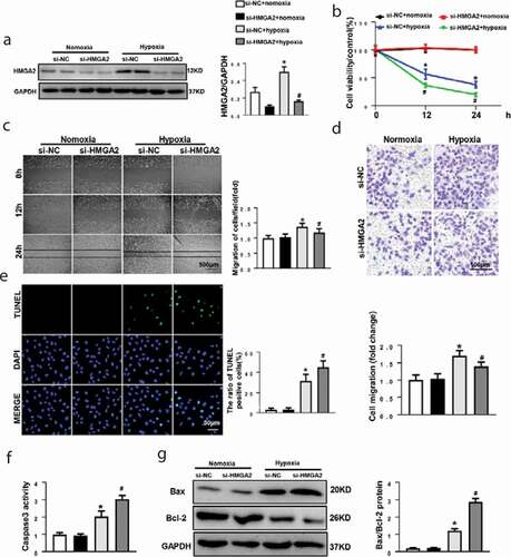
Figure 4. HMGA2 regulated HIF-1α/VEGF pathway
(a) Representative blots of HIF-1α, HIF-2α,VEGF,P-Erk1/2, eNOS and AKT expression of HUVECs after infection of Ad-GFP or Ad-HMGA2 vector with or without hypoxia stimulation for 12 h and all of the proteins were normalized to GAPDH(n = 6). (b) Effect of HMGA2 overexpression on VEGF release with hypoxia treatment for 12 h (n = 6). (C) NO production in the indicated groups under hypoxia treatment for 12 h (n = 6). (b–c): *P < 0.05 vs. Ad-GFP+nomoxia group. #P < 0.05 vs. Ad-GFP+hypoxia group. All quantitative data were presented as the mean ± SD. (d) Representative blots of HIF-1α, HIF-2α,VEGF, P-Erk1/2, eNOS and AKT expression of HUVECs after transfection of si-NC or si-HMGA2 with or without hypoxia treatment for 12 h and all of the proteins were normalized to GAPDH(n = 6). (e) Effect of HMGA2 overexpression on VEGF release with hypoxia treatment for 12 h (n = 6). (f) NO production in the indicated groups under hypoxia treatment for 12 h (n = 6). (e–f): *P < 0.05 vs. si-NC+nomoxia group. #P < 0.05 vs. si-NC+hypoxia group. All quantitative data were presented as the mean ± SD.
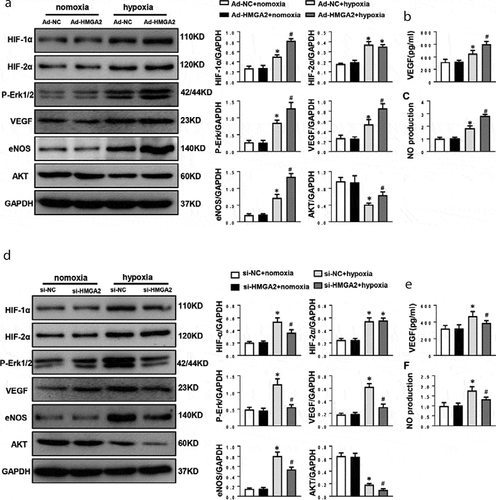
Figure 5. HMGA2 promoted migration by activating eNOS and inhibited apoptosis by regulating AKT
(a) Representative blots of the effects of si-VEGFR2, si-eNOS and si-AKT(n = 6). (b) The wound healing assay and the number of migrated cells in HUVECs in indicated group(n = 6). (c) Representative images and quantification of cell migration under hypoxia 24 h in Transwell chamber assay(n = 6). (d) TUNEL staining and quantitative analysis of apoptotic cells in HUVECs in indicated group under hypoxia for 12 h (n = 6). (b–d): aP < 0.05 vs. the control group. bP < 0.05 vs. the Hypoxia group. cP < 0.05 vs. the Ad-HMGA2+ hypoxia group. All quantitative data were presented as the mean ± SD.
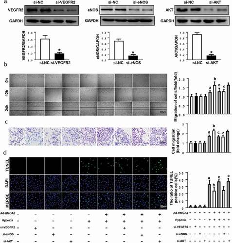
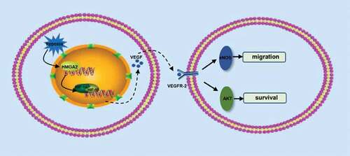
Figure 6. A putative scheme illustrating the mechanism by which HMGA2 contributes to pro-migration and anti-apoptosis of HUVECs in hypoxia-induced injury
Supplemental material