Figures & data
Figure 1. Expression pattern of USP1 and WDR48 during TGF-β stimulation. (a–b) USP1 and WDR48 expression in TNBC and non-TNBC breast cancer patients according to the data in METABRIC (a) and TCGA (b). (c–d) The relative expression of USP1 and WDR48 in different cell lines, including hormone receptor (HR) positive cell lines, M7 and T47D, and triple-negative cell lines, MDA-MB-231 and MDA-MB-468 (c–d). (e–h) RT-PCR analysis of the expression pattern of USP1 (e, g) and WDR48 (f, h) in MDA-MB-231 and MDA-MB-468 cells during TGF-β stimulation. (i–j) Western blot analysis of USP1 and WDR48 expression in MDA-MB-231 (i) and MDA-MB-468 (j) cells stimulated with TGF-β for the indicated time periods
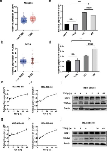
Figure 2. USP1/WDR48 complex enhanced TGF-β-induced migration and invasion in the TNBC cells. (a–b) Transwell migration and invasion assays for MDA-MB-231 (a) and MDA-MB-468 cells (b) treated with or without TGF-β and ML323. All representative images were obtained from at least three independent experiments
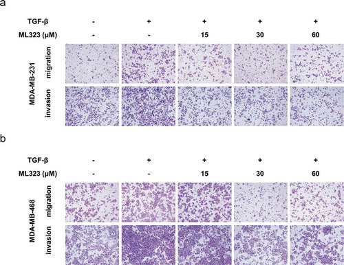
Figure 3. USP1/WDR48 complex promoted TGF-β-induced EMT. (a–b) MDA-MB-231 and MDA-MB-468 cells treated with or without TGF-β and ML323 for 12 h, the protein levels of EMT markers were analyzed by western blot. (c) The mRNA expression levels were analyzed by RT-PCR. (d–e) Western blot and RT-PCR analysis of USP1 (d) or WDR48 (e) expression in MDA-MB-231 and MDA-MB-468 cells transfected with the indicated siRNA for 48 h. (f–g) MDA-MB-231 and MDA-MB-468 cells were transfected with control siRNA, USP1 siRNA 2, or WDR48 siRNA 1 for 48 h and then stimulated with PBS or TGF-β for 24 h. Western blot analysis of the EMT markers expression pattern. All representative images were obtained from at least three independent experiments. *P < 0.05, **P < 0.01, *** P < 0.001, **** P < 0.0001 by Student’s t-test. Error bars are defined as s.d
Figure 4. USP1/WDR48 knockdown inhibited TGF-β-induced-Smad2/3 and MAPK activation. (a–d) MDA-MB-231 (a–b) and MDA-MB-468 (c–d) cells were transfected with control siRNA, USP1 siRNA 2, or WDR48 siRNA 1 for 48 h and stimulated with TGF-β for the indicated time periods. Western blot analysis of Jnk/p-Jnk, Erk/p-Erk, p38/p-p38, Smad2/p-Smad2, and Smad3/p-Smad3 expression
Figure 5. USP1/WDR48 complex directly binds to TAK1 and mediates its stability by downregulating its polyubiquitination. (a) Lysates from HEK293T cells transiently cotransfected with Flag-USP1 and Myc-Smad2, Myc-Smad4, Myc-TAK1, or Myc-TRAF6 were subjected to immunoprecipitation with anti-Flag antibody followed by Western blot analysis with anti-Myc antibody. (b) Lysates from HEK293T cells transiently cotransfected with Myc-TAK1 and Flag-WDR48 or Flag-USP1 were subjected to immunoprecipitation with anti-Myc antibody followed by Western blot analysis with anti-Flag antibody. (c) Lysates from MDA-MB-231 cells stimulated with TGF-β for indicated periods were subjected to immunoprecipitation with the TAK1 antibody or control IgG followed by Western blot analysis with the indicated antibodies. Protein in whole-cell lysate was used as positive control (Input). (d) Western blot analysis of extracts from MDA-MB-231 cells treated with 30 µM ML323 for 4 h and then stimulated with TGF-β for the indicated time periods. (e) Western blot analysis of lysates from HEK293T cells transfected with HA-tagged ubiquitin (HA-Ub), Myc-TAK1 and Flag-USP1, or Flag-WDR48, followed by immunoprecipitation with anti-Myc, probed with anti-HA