Figures & data
Table 1. Expression patterns of CHPF in cholangiocarcinoma tissues and normal tissues revealed in immunohistochemistry analysis
Table 2. Relationship between CHPF expression and tumor characteristics in patients with cholangiocarcinoma
Table 3. Relationship between CHPF expression and tumor characteristics in patients with cholangiocarcinoma analyzed by Spearman rank correlation analysis
Figure 1. CHPF was upregulated in cholangiocarcinoma
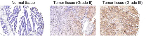
Figure 2. Construction of cholangiocarcinoma cell models with CHPF knockdown
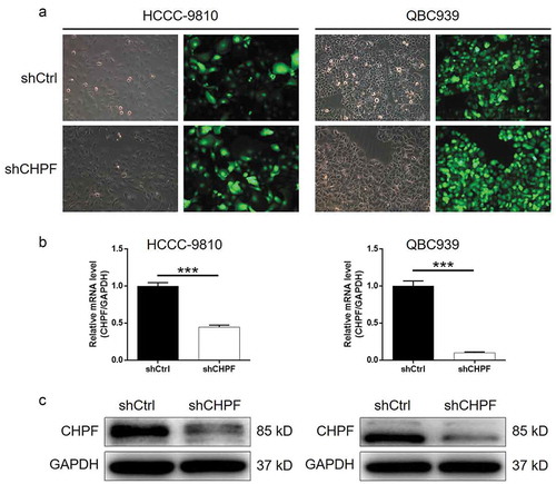
Figure 3. CHPF knockdown inhibited cell proliferation and promoted cell apoptosis and cell cycle arrest in cholangiocarcinoma cells
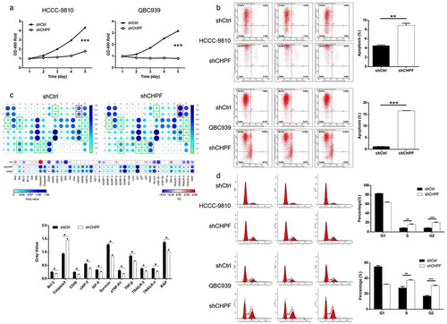
Figure 4. CHPF knockdown inhibited cell migration and expression of EMT-related proteins
Figure 5. CHPF overexpression promoted cholangiocarcinoma development in vitro.
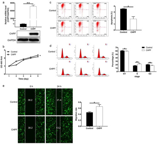
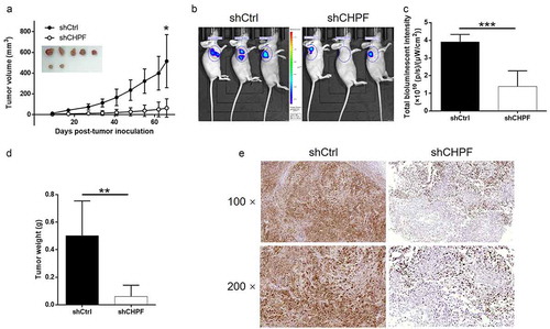
Figure 6. CHPF knockdown inhibited tumor growth of cholangiocarcinoma in vivo.