Figures & data
Table 1. Primers used in real-time PCR analysis
Figure 1. The identification of EXs around MSCs and the expression level of miR-145-5p were detected in MSC-EXs
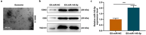
Figure 2. MSC-EXs (EX) containing miR-145-5p improved functional recovery and reduced histopathological injury in SCI rats
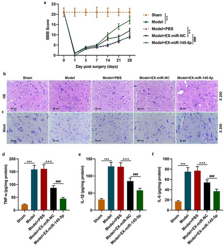
Figure 3. MSC-EXs could promote the expression of miR-145-5p in spinal cord tissue and inhibit the activation of TLR4/NF- κB pathway in SCI rats
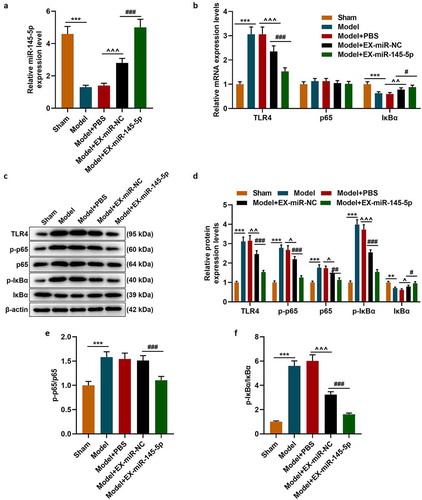
Figure 4. MSC-EXs could inhibit the effect of LPS inhibition ofon inhibiting miR-145-5p expression, cell viability and apoptosis in PC12 cells
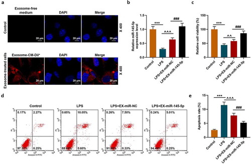
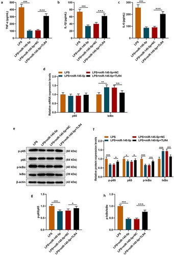
Figure 5. MSC-EXs inhibited LPS-induced inflammatory response and activation of the TLR4/NF-κB pathway in PC12 cells
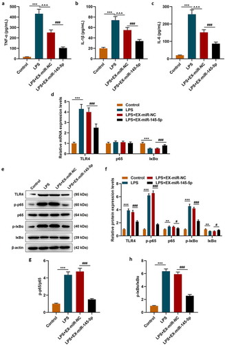
Figure 6. MiR-145-5p could specifically target TLR4 and inhibit TLR4 expression
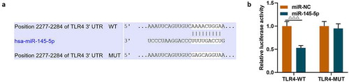
Figure 7. MiR-145-5p inhibited TLR4 expression and TLR4 overexpression significantly reversed the protective effect of EX-miR-145-5p on PC12 cell viability, inhibition of apoptosis
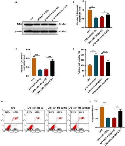
Figure 8. TLR4 overexpression significantly reversed the effect of EX-miR-145-5p on inhibiting inflammatory response and activating TLR4/NF- κB pathway in PC12 cells