
Figures & data
Figure 1. Knock down of MAGT1 inhibits proliferation and induces S-phase arrest and apoptosis in HeLa cells
(a) RT-qPCR analysis of mRNA levels of MAGT1 in three different siRNAs and negative control treated HeLa cells. Noting that SiMAGT1-1 and SiMAGT1-2 could significantly knock down MAGT1 gene. The results are presented as mean ± SD; n = 5; ***P < 0.001; ns, no significance; two-tailed Student’s t-test.(b) Western blot analysis of MAGT1 in different siRNAs treated HeLa cells. Quantification shown in right panel is from three independent experiments. The results are presented as mean ± SD; n = 3; **P < 0.01; ***P < 0.001; two-tailed Student’s t-test.(c) Representative pictures of the HeLa cells treated with SiCtrl, SiMAGT1-1 or SiMAGT1-2. The figures of high resolution showed the morphologic change of SiMAGT1 treated cells compared with negative control cells. Scare bars, 200 μm. (d) The number of different siRNAs treated cells counted through a cell counter. Noting that the cell number significantly reduced in MAGT1 RNAi groups at 48 h and 72 h compared with control. The results are presented as mean ± SD; n = 3; **P < 0.01; ***P < 0.001; ns, no significance; one-way ANOVA followed by Bonferroni’s multiple comparison tests. (e) Representative flow cytometry plots of cell cycle distribution propidium iodide (PI) staining in HeLa cells 72 hours after siRNAs treatment. Samples are measured by a flow cytometer with a minimum of 20,000 cells recorded from each experiment. Noting that MAGT1 RNAi changes the cell phase distribution of the HeLa cells. (f) Quantification of cell cycle analysis of PI staining in different siRNAs treated HeLa cells. Noting that MAGT1 RNAi significantly increased the cells in S-phase. The results are presented as mean ± SD; n = 4; *P < 0.05; ns, no significance; two-tailed Student’s t-test.(g) Representative flow cytometry plots of cell cycle analysis of SiCtrl and SiMAGT1 treated HeLa cells by EdU pulse-labeling and 7-AAD staining. Samples are measured by a flow cytometer with a minimum of 20,000 cells recorded from each experiment. (h) Quantification of the EdU positive cells in different siRNAs treated groups. Noting that MAGT1 RNAi did not significantly affect the EdU positive cells in different siRNAs treated groups. The results are presented as mean ± SD; n = 3; ns, not significant; two-tailed Student’s t-test. (i) Representative flow cytometry plots of apoptosis analysis of SiCtrl and SiMAGT1 treated HeLa cells by Annexin V and PI staining. Samples are measured by a flow cytometer with a minimum of 20,000 cells recorded from each experiment. (j) Quantification of apoptosis analysis of SiCtrl and SiMAGT1 treated HeLa cells. The results are presented as mean ± SD; n = 3; *P < 0.05; ns, no significance; two-tailed Student’s t-test.
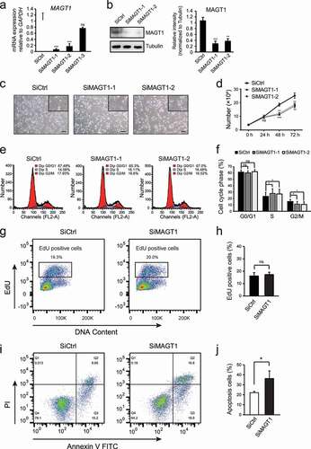
Figure 2. MAGT1 regulates several cell cycle related gene expressions
(a) RT-qPCR analysis of mRNA levels (relative to GAPDH) of MAGT1 in different siRNAs treated HeLa and SiHa cells. (b) RT-qPCR analyses of mRNA levels (relative to GAPDH) of CCNA1 and CCNB1 in different siRNAs treated HeLa and SiHa cells. (c) RT-qPCR analyses of mRNA levels (relative to GAPDH) of CCND1, CCNE1, CDK2 and CDK4 in different siRNAs treated HeLa and SiHa cells. (d) Western blot analysis of the protein level of the cycle-related regulators (cyclin-A1, cyclin-B1, cyclin-D1, cyclin-E1, CDK2, and CDK4). Tubulin is supported as the loading control. The black star indicates the specific target band. (e) RT-qPCR analyses of mRNA levels (relative to GAPDH) of CDKN2A and CDKN1A in different siRNAs treated HeLa and SiHa cells. (f) RT-qPCR analyses of mRNA levels (relative to GAPDH) of TP53 and MYC in different siRNAs treated HeLa and SiHa cells. For all the data above are shown as mean ± SD; n = 3; *P < 0.05; **P < 0.01; ***P < 0.001; ns, no significance; two-tailed Student’s t-test.
Figure 3. MAGT1 regulates a large batch of target gene expression
(a) Volcano Plots of differentially expressed genes (DEG) between cells treated with control and MAGT1 siRNAs, as determined by the RNA-seq analysis. 1,598 significantly changed genes are selected for further analyses, based on the indicated rules P < 0.05, |Log2 Fold change| >1. (b) Reactome Enrichment analysis of 1,598 MAGT1 regulated genes. (c) GO charts of Biological Processes, Cellular Component and Molecular Functions for 681 UP expressed genes in MAGT1 knock down group. (d) GO charts of Biological Processes, Cellular Component and Molecular Functions for 917 DOWN expressed genes in MAGT1 knock down group.
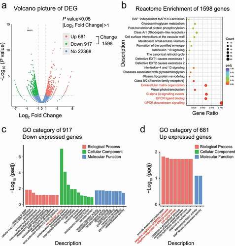
Figure 4. Downregulation of MAGT1 causes the reduction of ERK/p38 MAPK signaling pathways
(a) Heat maps of normalized expression values (Log2 unit) of RNA-seq data for genes in different RNAi-treated HeLa cells as indicated. The 14 genes indicated are negative regulators of MAPK cascade. (b) The Log2 fold change of MAPK pathway genes in different treated groups, noting that MAGT1 RNAi induced the up expression of 14 MAPK negative regulated genes, analyzed by KEGG. (c) Western blot analysis of the protein and phosphorylation level of the three MAPK core factors ERK, p38, and JNK. Tubulin is supported as the loading control.
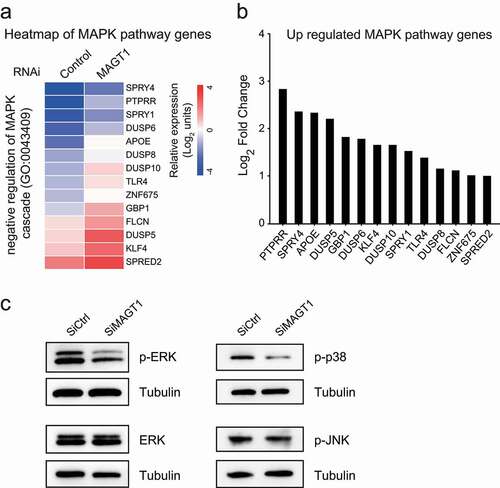
Figure 5. MAGT1 is required for E6/E7 proliferation regulation and G1/S transition
(a) The number of different siRNAs treated cells counted through a cell counter. Noting that cell number significantly reduces in MAGT1 plus E6/E7 RNAi group at 48 h and 72 h compared with E6/E7 RNAi group. The results are presented as mean ± SD; n = 3; **P < 0.01; ns, no significance; one-way ANOVA followed by Bonferroni’s multiple comparison tests. (b) Histogram showing cell cycle distribution of HeLa cells, 48 h and 72 h after SiRNA treatment. Noting that MAGT1 antagonizes E6/E7 RNAi-induced G0/G1 phase arrest in HeLa cells. (c) Effects of different RNAi on cell cycle distribution of HeLa cells. Samples are measured by a flow cytometer with a minimum of 20,000 cells recorded from each experiment. All results are presented as mean ± SD; n = 3; *P < 0.05; ***P < 0.001; ns, no significance; ##P < 0.01; ###P < 0.001; two-tailed Student’s t-test.
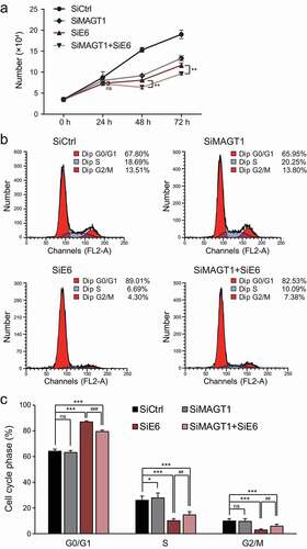
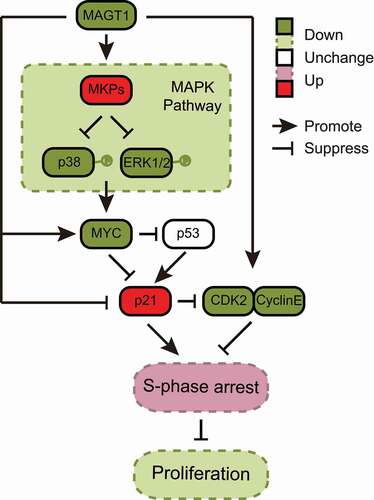
Figure 6. Molecular mechanism of MAGT1 in HeLa cell proliferation
Knocking down MAGT1 via p21 activation mediates S-phase arrest and inhibition of the ERK/p38 MAPK MYC signaling axis. Generally, knock down of MAGT1 causes the reduction of ERK/p38 MAPK signaling pathway, which might cause the downregulation of MYC. Low level of MYC could not antagonize p53 in p21 inhibition, thus leading to the remarkable upregulation of p21. The high level of p21 accompanied by downregulated CDK2 and cyclin-E ultimately induces S-phase arrest and the cell proliferation attenuation.
Data availability statement
The GEO accession number for the RNA-seq data is GSE166084. The data that support the findings of this study are available from the corresponding author upon reasonable request. https://www.ncbi.nlm.nih.gov/geo/query/acc.cgi?acc=GSE166084