
Figures & data
Figure 1. A comparison of hAECs isolation protocols. AM, amniotic membrane; Cells (m)/g, cells (in million) per gram of tissue; cells (M)/amnion, cells (in million) per amnion; Cg, collagenase; CF, calcium-free; CHM, cell heterogeneity markers; CMF, calcium magnesium-free; CM, clinical method; ESCM, epithelial cell surface markers; GS, gentle shaking; IM, immunogenicity markers; NA, not available; P/S, penicillin/ streptomycin; PM, pluripotency markers; SCM, stem cell markers for non-differentiated cells; SW, shaking water bath; TM, traditional method; VS, vigorous shaking. *Original protocols.
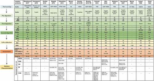
Table 1. A comparison of hAECs culture protocols.
Table 2. Commercially available human amniotic epithelial cell lines.
Table 3. Comparison of cryopreservation media.
Table 4. Clinical trials using hAECs as a method of intervention.