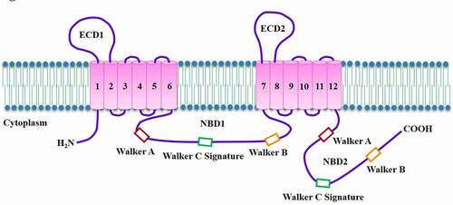
3,173
Views
10
CrossRef citations to date
0
Altmetric
Review

Molecular mechanisms for ABCA1-mediated cholesterol efflux

, , , &
Pages 1121-1139 | Received 04 Jan 2022, Accepted 24 Jan 2022, Published online: 22 Feb 2022

Figures & data

Figure 1. Topological diagram of ABCA1.

ABCA1 is a full transporter that is composed of two ECDs, multiple membrane-spanning segments, 12 TMs, and two NBDs. Each NBD contains Walker A, Walker B, and Walker C signature.
Figure 1. Topological diagram of ABCA1.

Table 1. The biological functions of ABCA1

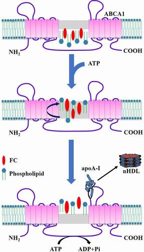
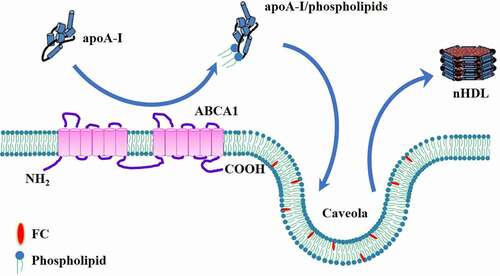
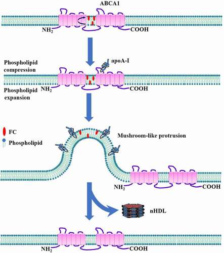
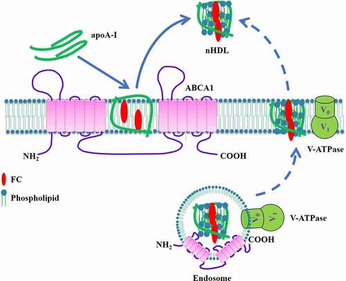
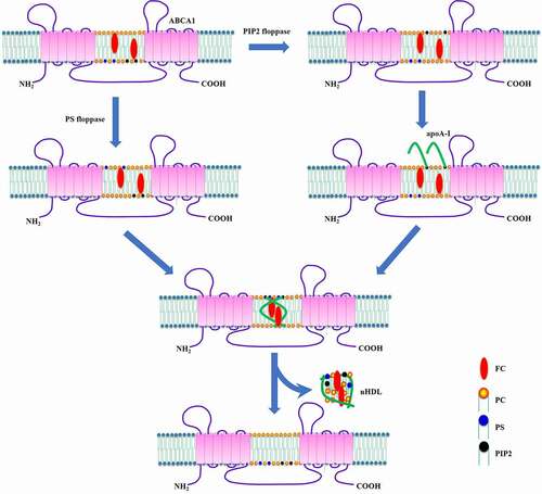
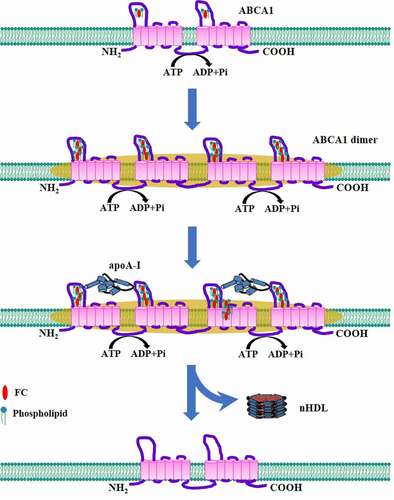
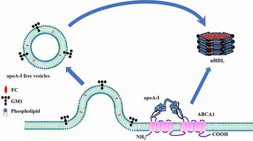
Table 2. The proposed models to explain ABCA1-mediated cholesterol efflux

Figure 2. Schematic illustration of the channel trafficking model.

Accumulated phospholipids along with FC in the inner leaflet of plasma membrane are laterally delivered to the chamber between both TMDs of ABCA1. ATP binding promotes NBD dimerization and closes the chamber, leading to lipid translocation to the outer leaflet. Subsequently, ATP is hydrolyzed to open the chamber at the top, which is followed by lipid export to the elongated hydrophobic tunnel. The hydrolysis of ATP also promotes apoA-I binding to ABCA1 for nHDL biogenesis. The chamber returns to the initial structure to absorb membrane lipids again after ADP dissociates from the NBDs.
Figure 2. Schematic illustration of the channel trafficking model.

Figure 3. Schematic illustration of two-step process model.

ABCA1 first promotes the translocation of phospholipid molecules to apoA-I. The phospholipid/apoA-I complex then picks up FC in the caveola in an ABCA1-independent manner, leading to nHDL formation.
Figure 3. Schematic illustration of two-step process model.

Figure 4. Schematic illustration of mushroom-like protrusion model.

Upon binding to a small amount of apoA-I, ABCA1 translocates FC and phospholipids from the inner leaflet to the outer leaflet of plasma membrane. This process leads to the lateral compression of phospholipid molecules in the outer leaflet and their expansion in the inner leaflet. Due to the unequal molecular packing density across the membrane, the mushroom-like protrusions are formed to release surface tension. A large amount of apoA-I molecules then bind to these protrusion structures. After accepting enough lipids, apoA-I dissociates from plasma membrane, leading to nHDL generation and membrane structure restoration.
Figure 4. Schematic illustration of mushroom-like protrusion model.

Figure 5. Schematic illustration of V-ATPase-induced apoA-I unfolding model.

ABCA1 recruits V-ATPase to the cell surface. V-ATPase then acidifies apoA-I and plasma membrane to unfold its N-terminus and enhance lipid fluidity, respectively. After partial unfolding, apoA-I forms the dimers that can spontaneously interact with membrane lipids, leading to the generation and release of nHDL. The dashed line indicates that V-ATPase may also contribute to the acidification and unfolding of apoA-I in the endosomes, followed by retroendocytosis and nHDL secretion.
Figure 5. Schematic illustration of V-ATPase-induced apoA-I unfolding model.

Figure 6. Schematic illustration of PIP2-induced apoA-I unfolding model.

ABCA1 flops PS and PIP2 to the outer leaflet where PIP2 interacts with apoA-I. This leads to apoA-I unfolding at its N-terminal domain and dimerization. The apoA-I dimer then inserts into plasma membrane to form nHDL.
Figure 6. Schematic illustration of PIP2-induced apoA-I unfolding model.

Figure 7. Schematic illustration of ABCA1 dimerization model.

The ABCA1 monomers translocate FC and phospholipids from plasma membrane to their ECDs in an ATP-dependent manner. When enough lipid molecules are sequestered by the ECDs, these ABCA1 monomers are converted to the dimers. Lipid-free apoA-I then binds to the ECDs of dimerized ABCA1. Lipids are loaded onto apoA-I, leading to nHDL generation. The ABCA1 dimers dissociate into monomers to transport lipids again.
Figure 7. Schematic illustration of ABCA1 dimerization model.

Figure 8. Schematic illustration of apoA-I-free vesicle model.

In the absence of apoA-I, FC and phospholipid molecules are transferred by ABCA1 to the outer leaflet leading to the formation of mushroom-like protrusions, which are released into the extracellular space as apoA-I-free vesicles. Upon binding to ABCA1, apoA-I unfolds at its N-terminal domain and then dimerize. The apoA-I dimers receive FC and phospholipids to form small nHDL. Lipids, including GM1, within apoA-I-free vesicles are subsequently loaded onto nHDL to generate larger particles.
Figure 8. Schematic illustration of apoA-I-free vesicle model.

Figure 9. Schematic illustration of sequential apoA-I addition model.

Each nHDL particle is derived from a separate step in the process of lipidation. Binding of one free-lipid apoA-I molecule to ABCA1 at plasma membrane leads to the production of poor-lipid apoA-I (<6 nm). If two free-lipid apoA-I molecules interact with ABCA1, 7.5 nm nHDL is formed. However, 10 ~ 12 nm nHDL is generated when three free-lipid apoA-I molecules bind to ABCA1.
Figure 9. Schematic illustration of sequential apoA-I addition model.

Figure 10. Schematic illustration of the retroendocytosis model.

ApoA-I interacts with cell surface ABCA1 to form a complex that is endocytosed to endosomal compartments via a Rab5-dependent pathway. ABCA1 translocates lipids to apoA-I for nHDL production. Rab4 promotes internalized ABCA1 and synthesized nHDL back to the cell surface. The nHDL particles are then released into the extracellular space, and ABCA1 is located in plasma membrane again.
Figure 10. Schematic illustration of the retroendocytosis model.

Reprints and Corporate Permissions

Please note: Selecting permissions does not provide access to the full text of the article, please see our help page How do I view content?

To request a reprint or corporate permissions for this article, please click on the relevant link below:

Academic Permissions

Please note: Selecting permissions does not provide access to the full text of the article, please see our help page How do I view content?

Obtain permissions instantly via Rightslink by clicking on the button below:

If you are unable to obtain permissions via Rightslink, please complete and submit this Permissions form. For more information, please visit our Permissions help page.