
Figures & data
Figure 1. Phosphorylation of CypA at Ser77 regulates subcellular localization.
A, Whole cell lysates from peripheral blood mononuclear cells (PBMC) and malignant haematological cell lines were resolved by SDS-PAGE and Western Blots were probed with anti-CypA primary antibody. Membranes were re-probed with anti-beta-actin as a loading control. B, Whole cell extracts of K562 cells were collected prior and post-release from a double-thymidine block (2 mM) followed by RO-3306 (9 µM) treatment (as outlined in below), and examined by SDS-PAGE and Western Blot using anti-CypA, anti-Cyclin B1, anti-Nek2 and anti-GAPDH primary antibodies. Uncropped blots can be found as part of the Supplementary Information. C, Densitometry plots highlighting changes in Cyclin B1 and Nek2 expression levels observed in whole cell extracts from B following release from synchronization. D, Whole cell extracts of K562 cells were collected and incubated at 30°C and 37°C for 30 min, in the presence or absence of Lambda phosphatase. Membranes were probed with anti-CypA and anti-GAPDH primary antibodies. E, F, G, K562 cells were transfected with pEGFP, pEGFP-CypAWT (E and F), pEGFP-CypAS51A, pEGFP-CypAS77A, pEGFP-CypAT93A (E), or pEGFP-CypAS51E, pEGFP-CypAS77E, pEGFP-CypAT93E (F). Cells were prepared on slides 48 hr post-transfection and stained with anti-pericentrin (red) and DAPI (blue). Image contrast was adjusted post-acquisition to enhance centrosome visualization. G, Centrosome and midbody localization of EGFP-CypAS77E and EGFP-CypAS77A was examined. Cells were transfected as indicated, with interphase and cytokinetic populations enriched and prepared on slides. Interphase cells were stained with anti-pericentrin (red), while cytokinetic cells were stained with anti-alpha-tubulin (red). All cells were counter-stained with DAPI. Bar graphs presented show the qualitative assessment of localization to the centrosome or midbody for EGFP-CypAS77E and EGFP-CypAS77A relative to EGFP-CypAWT (n=30 for each protein at both localizations), as well as the quantification of changes in signal intensity at the centrosome and midbody relative to EGFP-CypAWT as well as. In the case of the latter, signal intensity at the centrosome was quantified for all cells examined, including those with apparent CypA mislocalization. Arrowheads indicate location of the centrosome (E, F) and midbody (G). Scale bar: 10 µm.
Please check axis of fig 1C axis title is masked - ”R” in ”Relative”
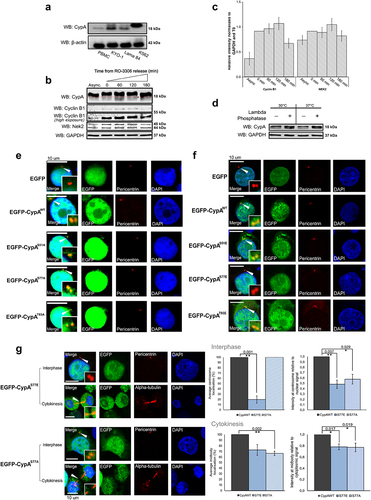
Figure 2. CypA interacts with Nek2 and PP1 during the cell cycle.
A, Schematic of K562 cell treatment to enrich at G2/M boundary and sample collection during mitotic progression. B, Cell cycle distribution of K562 cells at T=0 and upon release from RO-3306, as measured by flow cytometry with staining of DNA content using propidium iodide. Graph represents mean data from 3 independent experiments, determined using the Multicycle Analysis feature on De NovoTM FCS Express software. C, Whole cell lysates from samples collected post-release from RO-3306 were resolved by SDS-PAGE and subjected to Western Blot analysis. The membranes were probed with anti-Nek2 and anti-Cyclin B1 primary antibodies. D, F, K562 cells were transfected with pEGFP and pEGFP-CypAWT, lysed 48 hr post-transfection and exogenous CypA was immunoprecipitated using GFP-Trap® beads and resolved by SDS-PAGE. Resolved proteins were analyzed by Western Blot using anti-Nek2 (D) and anti-PP1 (F) primary antibody. E, G, K562 cells were transfected with pEGFP and pEGFP-CypAWT and synchronised to the G2/M border as indicated in A. Cells were collected 60 min post-release from RO-3306, and CypA was immunoprecipitated and analyzed by SDS-PAGE and Western Blot. Membranes were probed with anti-Nek2 (E) and anti-PP1 (G) primary antibody. Whole-cell extracts (WCE) and post-binds (P-B) collected after immunoprecipitation in D-G were probed with anti-GFP to confirm the presence and absence of exogenous EGFP.
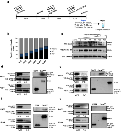
Figure 3. CypA is phosphorylated by Nek2 in vitro.
A, Recombinant active Nek2 or kinase-inactivated Nek2 (KD-Nek2) were incubated alone, with purified Casein or His-CypAWT in the presence of [γ-32P]-labelled ATP for 60 min at 30°C. KD-Nek2 was generated by heating in 5 mM DTT for 5 min at 95°C. The incorporation of radio-labelled ATP was examined by autoradiography following SDS-PAGE and transfer onto PVDF membranes. Once the [γ-32P]-ATP had sufficiently decayed, membranes were probed with anti-Nek2 and anti-CypA. B, Purified His-CypAWT, His-CypAS51A, His-CypAS77A and His-CypAT93A were incubated with [γ-32P]-ATP in the presence and absence of active recombinant Nek2 for 60 min at 30°C and the incorporation of radioactive ATP was examined by autoradiography following SDS-PAGE and transfer onto PVDF membranes. Membranes were probed with anti-Nek2 and anti-CypA primary antibodies after sufficient decay of the [γ-32P]. C, Quantification of total [γ-32P]-ATP incorporation by each recombinant protein relative to corresponding Nek2 autophosphorylation levels observed through densitometry. Quantification was further separated into [γ-32P]-ATP incorporation by the upper and lower bands indicated in B, that represent phosphorylated forms of CypA with differing mobilities. Graph represents mean ± SEM of three independent experiments (p-value < 0.05). D, Chymotrypsin cleavage of the substrate N-succinyl-Ala-Ala-Pro-Phe-pNitroanilide was quantified in absence of CypA (CypA-free), or presence of His-CypAWT, His-CypAS51A, His-CypAR55A, His-CypAS77A and His-CypAT93A. Absorbance of pNitroaniline was measures at 390 nm. Results shown are mean values obtained for each time point across three independent experiments.
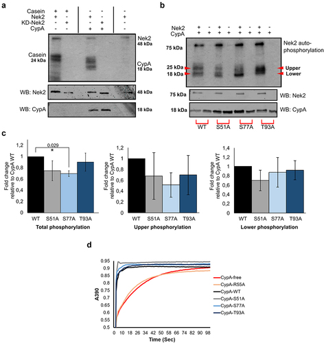
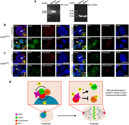
Figure 4. CypA does not affect Nek2 or PP1 localization to the midbody.
A, Homologous knock-out of CypA in JurkatCypA-/- cells was confirmed by Western Blot with anti-CypA, and comparison to wild-type Jurkat cells (JurkatCypA+/+). GAPDH was used as a loading control. B, C, Interphase cells were prepared on slides and stained with anti-Nek2 (green), anti-PP1 (green), anti-pericentrin (red) and DAPI (blue). Cytokinetic cell populations were generated by treatment with nocodazole (160 nM) for 16 hr, washed in PBS and released into complete media for 75 min (JurkatCypA+/+) and 90 min (JurkatCypA-/-) before slide preparation. Cells were stained with anti-Nek2 (green), anti-PP1 (green), anti-alpha-tubulin (red) and DAPI (blue). Arrowheads indicate location of the centrosome and midbody (B, C). Scale bar: 10 µm. D, CypA may exist as part of a Nek2-PP1 complex. During mitosis, Nek2 expression is elevated and PP1 is lost from the complex, which together increase Nek2 activity towards CypA, triggering centrosome dissociation and midbody recruitment during cytokinesis.
Replace above sentence D ”CypA may exist as part of novel component of a Nek2-PP1 complex” with ”CypA may exist as part of a Nek2-PP1 complex”.
Supplemental material
Supplemental Material
Download Zip (22.6 MB)Data availability statement
The authors confirm that the data supporting the findings of this study are available within the article and/or its supplementary materials.