
Figures & data
Figure 1. IKKβ knockout accelerates senescence in mouse skin fibroblasts.
(a)(b) Sm22αCre-mediated recombination occurred spontaneously under standard culture conditions. Skin explants from adult IKKβFL/FL and IKKβFL/FLSm22αCre mice (n = 2) (a) and ROSAmT/mG Sm22αCre reporter mice (n = 2) (b) were plated on plastic dishes and cultured in DMEM supplemented with 10% FBS under 3% O2 and 5% CO2 conditions. The protein levels of IKKβ and GAPDH (the internal control) at passage 2 were measured by Western blotting (a) and the expression of EGFP and tdTomato was examined by fluorescence microscopy (b). Scale bars, 100 µm. (c)(d) Growth curves of skin fibroblasts under 20% and 3% O2 conditions, respectively (n = 5). Primary skin fibroblasts isolated from five adult IKKβFL/FL and IKKβFL/FLSm22αCre mice were cultured under physiological oxygen level (3-5% oxygen) conditions until passage 3 (P3), after which time, they were passaged in parallel according to a 3T2 protocol under 20% O2 conditions (c) or maintained under physiological oxygen level (3% oxygen) conditions according to a 3T1 protocol (d). The graph shows the cumulative number of cells in sequential passages. (e) Representative images showing senescence-associated β-galactosidase (SA-β-gal) staining of IKKβFL/FL and IKKβFL/FLSm22αCre cells cultured as in (c) (P7: 5 passages under 20% oxygen conditions). Scale bars, 100 µm. (f) Quantification of SA-β-gal staining of skin fibroblasts harvested during the indicated passages. Error bars represent the standard error of the mean. (*p value < 0.05, **p value < 0.01)
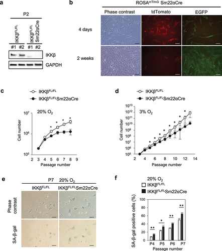
Figure 2. Expression of constitutively active IKKβ bypasses cellular senescence.
(a) Control (R26StopFLIKK2CA Cre-negative) and IKKβ-CA (R26StopFLIKK2CA Sm22αCre) skin fibroblasts were treated with MG132 (10 μM), a proteasome inhibitor, for 30 min prior to stimulation with IL-1β (10 ng/ml) for 5 min. Whole-cell lysates were analyzed by Western blotting using the indicated antibodies. Representative images are shown (n = 3). (b) Nuclear and cytoplasmic fractions were prepared from control and IKKβ-CA skin fibroblasts and analyzed by Western blotting. Nuclear p65 levels were quantified by ImageJ and normalized to the level of Lamin A (n = 3). (c)(d) Growth curves of control and IKKβ-CA skin fibroblasts under 20% and 3% oxygen conditions (n = 5). P2 Skin fibroblasts isolated from five adult control and IKKβ-CA mice were passaged in parallel under 20% oxygen conditions according to a 3T3 protocol and under 3% oxygen conditions according to a 3T1 protocol. The graph shows the cumulative number of cells in sequential passages. (e)(f) Representative images and quantification of senescence-associated β-galactosidase (SA-β-gal) staining of control and IKKβ-CA cells in passage 8 cultured under 3% and 20% oxygen conditions (n = 5). Scale bars, 100 µm. Error bars represent the standard error of the mean. (*p value < 0.05, **p value < 0.01)
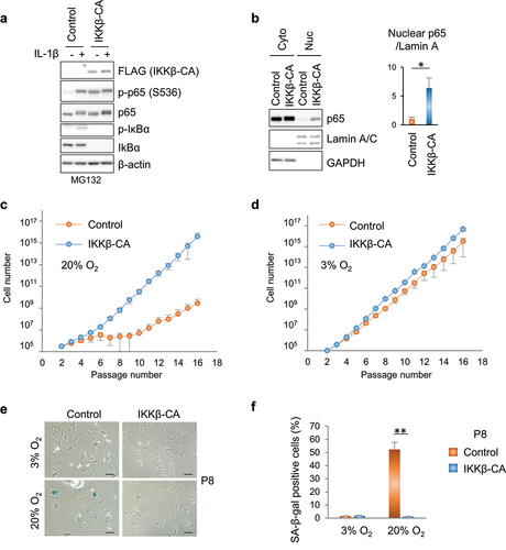
Figure 3. Expression of nondegradable IκBα abolishes IKKβ-CA-induced senescence bypass.
(a) Control (R26StopFLIKK2CA Cre-negative) and IKKβ-CA (R26StopFLIKK2CA Sm22αCre) skin fibroblasts were infected with lentivirus carrying an empty vector (EV) or FLAG-tagged IκBαSR. The expression of IκBαSR was confirmed by Western blotting using an antibody against the DYKDDDDK Tag. Representative images are shown (n = 4). (b) Nuclear and cytoplasmic fractions were prepared from control and IKKβ-CA skin fibroblasts harboring EV or IκBαSR. The nuclear p65 and p50 levels were measured by Western blotting, with quantification performed with ImageJ and normalized to the level of Lamin A (n = 4). (c)(d) Growth curves of control and IKKβ-CA skin fibroblasts harboring EV and IκBαSR cultured under 20% and 3% oxygen conditions, respectively. Skin fibroblasts were cultured under physiological oxygen level (3-5% oxygen) conditions until passage 3, at which time, they were infected with lentivirus. The cells were passaged again and treated with 2 μg/ml puromycin for 3 days under 3% oxygen conditions. The selected cells were passaged under 20% oxygen conditions according to a 3T2 protocol (c) or maintained under physiological oxygen level (3% oxygen) conditions according to a 3T1 protocol (d). The graph shows the cumulative number of cells in sequential passages. (e) Representative images and quantification of senescence-associated β-galactosidase (SA-β-gal) staining of control and IKKβ-CA cells harboring EV or IκBaSR cultured under 20% oxygen conditions. Scale bars, 100 µm. Error bars represent the standard error of the mean. (*p value < 0.05, **p value < 0.01)
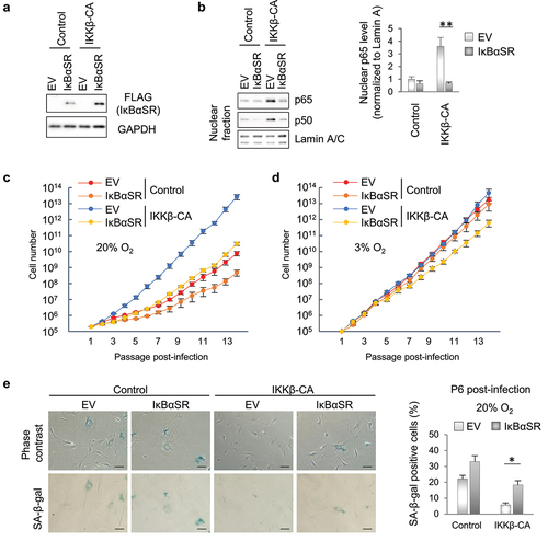
Figure 4. IKKβ-CA cells bypass senescence with functional p53 and normal levels of p21.
(a) (b) Control (R26StopFLIKK2CA Cre-negative) and IKKβ-CA (R26StopFLIKK2CA Sm22αCre) skin fibroblasts derived from four mice which had been maintained under 3% oxygen conditions until P3 were passaged under 20% oxygen conditions according to a 3T2 protocol. The cells were harvested during the indicated passages. p53 and p21 levels in whole cell extracts were analyzed by Western blotting (a). The nuclear fraction was extracted during passages 4 and 6, and analyzed by Western blotting (b). Representative images are shown. The nuclear p53 levels were quantified by ImageJ and normalized to the Lamin A/C levels (n = 4). (c) Control and IKKβ-CA skin fibroblasts were cultured as in (A). The mRNA levels of p53 target genes were measured by RT-qPCR and normalized to the level of GAPDH. (d) Control and IKKβ-CA skin fibroblasts derived from four mice were cultured as described in until passage 20, at which time they were treated with 500 nM doxorubicin (DoxR) for 4 hours. The p53 and p21 levels were measured by Western blotting. Error bars represent the standard error of the mean. (*p value < 0.05, **p value < 0.01)

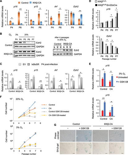
Figure 5. IKKβ suppresses the derepression of the genes in the INK4/Arf locus and counteracts the senescence-associated downregulation of Ezh2.
(A)(B)(C) IKKβ-CA and control cells (n = 4) which had been maintained under 3% oxygen conditions until P3 were passaged under 20% oxygen conditions according to a 3T2 protocol (a)(b). IKKβ-CA and control skin fibroblasts harboring an empty vector (EV) or IκBαSR were generated and passaged as described in . The mRNA levels of p16INK4a, p15INK4b, Arf and Ezh2 were measured by RT-qPCR and normalized to the geometric mean of GAPDH and TMEM199 expression levels (a)(c). Ezh2 levels in whole-cell extracts were analyzed by Western blotting (b). (d) IKKβFL/FLSm22α-Cre and IKKβFL/FL cells (n = 5) which had been maintained under 3% oxygen until P3 were passaged according to a 3T2 protocol under 20% oxygen conditions. The mRNA levels of p16INK4a and Ezh2 were measured by RT-qPCR at the indicated passages and normalized to the geometric mean of GAPDH and TMEM199 expression levels. (e) After 5 days of treatment with 5 μM GSK126, an Ezh2 inhibitor, under 3% oxygen conditions, total RNA was isolated from IKKβ-CA and control fibroblasts and the mRNA levels of p16INK4a and p15INK4b were measured by RT-qPCR and normalized to the levels of 18S rRNA. (f) Control and IKKβ-CA cells were serially passaged in the presence or absence of 5 μM GSK126 in 20% and 3% oxygen. (g) Representative images showing senescence-associated β-galactosidase (SA-β-gal) staining in control and IKKβ-CA cells treated with or without 5 μM GSK126 for 5 days (n = 4). Scale bars, 100 µm. Error bars represent the standard error of the mean. (*p value < 0.05; **p value < 0.01)
Supplemental material
Supplementary materials.pdf
Download PDF (2.1 MB)Data availability statement
The authors confirm that the data supporting the findings of this study are available within the article [and/or] its supplementary materials.