Figures & data
Figure 1 CT scan during baseline evaluation demonstrated mild-to-moderate hilar adenopathy with early calcification of lymph nodes. Mild air trapping with moderate bronchial wall thickening was noted. Linear band of opacity and ground galss opacities were noted in the right lower lobe. Minimal emphysema was reported in the right upper lobe.
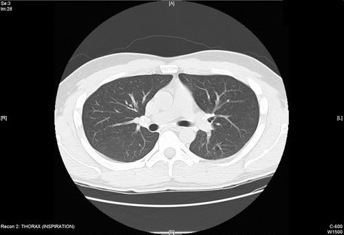
Figure 2 Follow up CT scan 3 months later while still exposed shows progressive bronchial wall thickening and development of new centriloblar nodules in the right upper lobe.
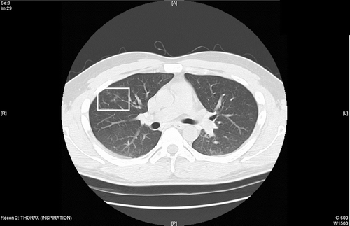
Figure 3 H & E stain of VATS biopsy demonstrates airways with a patchy mild chronic inflammatory infiltrate and interstitial macules that are perivascular and peribronchiolar in distribution associated with mild collagen deposition. Finely granulated macrophages within the interstitium were reported as consistent with aluminum pneumoconiosis.
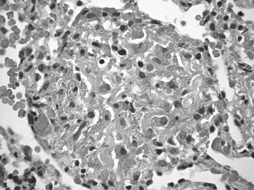
Table 1 Serial pulmonary function tests