Figures & data
Table 1 Baseline characteristics
Figure 1 Kaplan–Meier analysis showing that survival is greater following LVRS than following transplantation (p = 0.01) by the log-rank test.
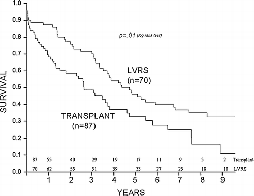
Table 2 1-year postoperative data of surviving patients
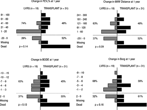
Figure 2 Compares the changes in FEV1, 6-minute walk test, mBODE and Borg from before surgery to 1-year post-surgery between LVRS patients (shown on the left) and transplantation patients (shown on the right). More transplantation patients are missing or dead at 1-year (below dotted line), while more LVRS patients did not improve following surgery (also below dotted line). Of patients who survived 1 year post-surgery, the transplantation patients had greater improvements in functional status (above dotted line).
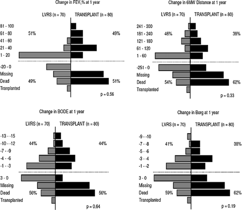
Table 3 Secondary outcomes
Figure 3 Causes of death in transplantation patients included complications from immunosuppression, medications, and acute and chronic graft failure.
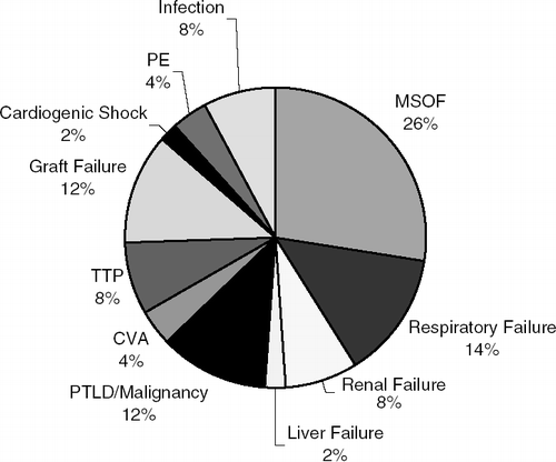
Figure 4 Kaplan–Meier analysis showing that survival is greater following LVRS than following transplantation in patients with baseline FEV1 20–30% at all times with a p = 0.04 by the log-rank test.
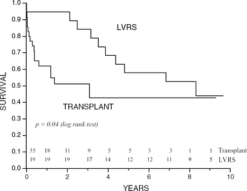
Figure 5 Compares changes in FEV1, 6-minute walk distance, mBODE and Borg from before surgery to 1-year post-surgery between LVRS (shown on the left) and transplantation patients (shown on the right) with a baseline FEV1 20–30%. More transplantation patients are missing or dead at 1-year (below dotted line). While no LVRS patients died within 1 year following surgery, more did not improve following surgery (also below dotted line). Of patients who survived 1 year post-surgery, the transplantation patients had greater improvements in functional status (above dotted line).