Figures & data
Figure 1 Cartoon of lung and chest wall with elastic tension represented by arrows. The difference between the tension of the two structures is the pleural pressure.
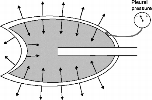
Figure 2 Graph of single breath interrupted technique for determining lung compliance. The subject slowly inspired from residual volume (RV) to total lung capacity and then expired back to RV. At several levels of the vital capacity maneuver, a shutter momentarily interrupts flow to eliminate the effect of airway resistance and diminish the hysteresis. After Forster et al. (Citation[15]), Figure 25.
![Figure 2 Graph of single breath interrupted technique for determining lung compliance. The subject slowly inspired from residual volume (RV) to total lung capacity and then expired back to RV. At several levels of the vital capacity maneuver, a shutter momentarily interrupts flow to eliminate the effect of airway resistance and diminish the hysteresis. After Forster et al. (Citation[15]), Figure 25.](/cms/asset/4fa3810a-7642-4879-a8fc-cf2b65640681/icop_a_216878_uf0002_b.gif)
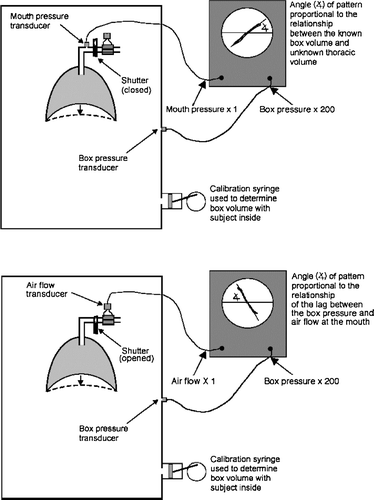
Figure 3 Simplified schematic diagrams of a body plethysmograph set up to measure lung volume (upper panel) and airway resistance (lower panel). See text for explanation.