Figures & data
Table 1 Bronchoalveolar lavage cell counts
Figure 1 Roflumilast reverses CS-induced downward trend of CCSP in BAL fluid. A: Representative image of CCSP Western blotting. Air: air control; CS: cigarette smoke alone; R+CS: roflumilast plus CS; R: roflumilast alone. B: Densitometry analysis of CCSP. Data are expressed as mean ± SEM normalized to the average CCSP protein level of air control group. n = 12–14 per group. *: p < 0.05 versus CS alone.
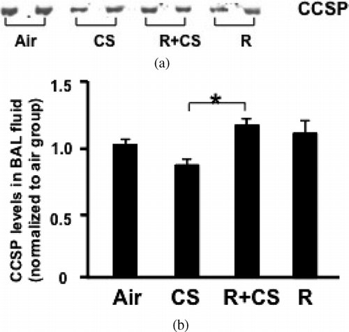
Figure 2 Roflumilast suppresses CS-induced upward trend of ERK1/2 activation in lungs. A: Representative image of ERK1/2 Western blotting. Air: air control; CS: cigarette smoke alone; R + CS: roflumilast plus CS; R: roflumilast alone. B: ERK1/2 activation was assessed by the ratio of p-ERK1/2 to t-ERK1/2. Densitometry analysis of ERK1/2 activation is presented in the chart diagram. Data are expressed as median (25%–75% range) normalized to the median ERK1/2 activation of air control group. n = 5–6 per group. *:p < 0.05 versus CS, #: p < 0.05 versus Air, CS, and R + CS.
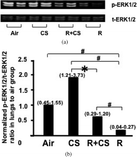
Figure 3 The activation level of ERK1/2 in lung tissues negatively correlated with CCSP levels of BAL fluid in CS alone, and roflumilast plus CS groups. The phosphorylation levels of ERK1/2 in lungs were plotted against CCSP levels in BAL fluid. The activation of ERK1/2 in lungs negatively correlated with CCSP levels of BAL fluid. (r2 = 0.45, p = 0.02).
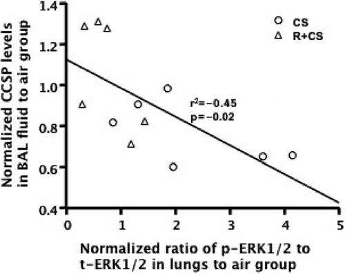
Figure 4 CS and roflumilast increase PCNA expression in airway epithelium. PCNA expression in airway epithelial cells was detected by immunohistochemistry in paraffin-embedded lung sections. (A) Representative lung photomicrographs of PCNA immunostaining. Air: air control; CS: cigarette smoke alone; R+CS: roflumilast plus CS; R: roflumilast alone. Arrows indicate positive cells. Internal scale bar = 50 μ m. (B) PCNA expression was assessed as number of positive epithelial cells per mm airway basement membrane. Values are presented as mean± SEM. *: p < 0.05 versus Air. #: p < 0.05 versus CS.
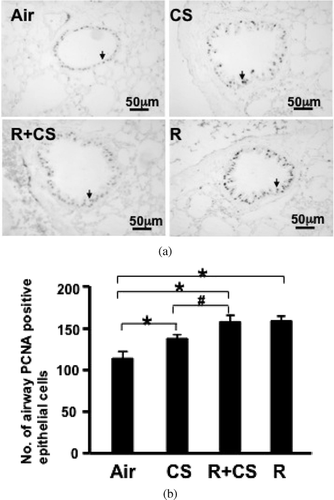
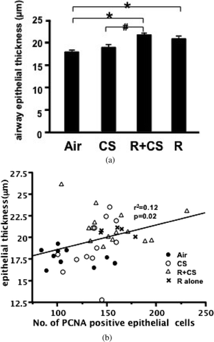
Figure 5 Roflumilast increases thickness of airway epithelium. (A) The airway epithelial thickness was calculated by dividing the epithelial area by the perimeter of corresponding basement membrane. Air: air control; CS: cigarette smoke alone; R+CS: roflumilast plus CS; R: roflumilast alone. Values are presented as mean ± SEM. *: p < 0.05 versus Air. #: p < 0.05 versus CS. (B) Airway epithelial PCNA level was plotted against the epithelial thickness. The airway PCNA expression positively correlated with the epithelial thickness (r2 = 0.12, p = 0.02).