Figures & data
Figure 1. Coronal images at maximum expiratory level (A, phase 20%) and maximum inspiratory level (B, phase 60%) at the identical anatomical position showing the diagnostic quality of the data.
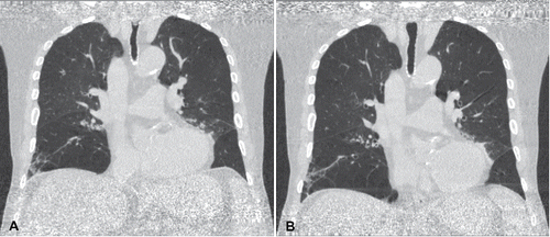
Figure 2. Changes of lung volume (A), emphysema volume (B) and emphysema index (C) over the whole respiratory cycle for each patient. One respiratory cycle (0–90%) was acquired and data were duplicated (100–190%) for better graphical visualization of the respiratory changes.
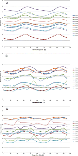
Table 1. Individual results for the range and mean () of lung volumes, mean lung densities, emphysema volumes and emphysema indices over the respiratory cycle.
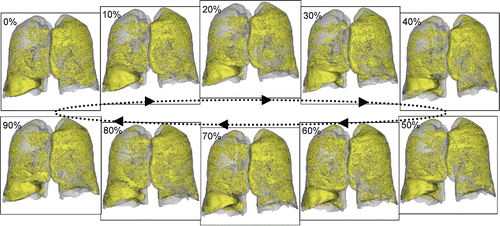
Figure 3. Segmentation results and visualization of the whole lung and the emphysematous areas (in yellow) during the whole respiratory cycle (from 0% to 90%). The lung volume in detail was: 0% – 5889 ml, 10% – 5777 ml, 20% – 5714 ml, 30% – 5754 ml, 40% – 5980 ml, 50% – 6251 ml, 60% – 6359 ml, 70% – 6313 ml, 80% – 6079 ml, 90% – 5963 ml. The emphysema index in detail was: 0% – 20.2%, 10% – 20.1%, 20% – 20.0%, 30% – 20.2%, 40% – 21.1%, 50% – 22.3%, 60% – 22.8%, 70% – 22.6%, 80% – 22.1%, 90% – 21.5%.