Figures & data
Table 1. Knowledge gaps and considerations for future studies.
Figure 1. Obese (OB) subjects with chronic obstructive pulmonary disease (COPD) (solid squares) had a rightward shifted dyspnea/ventilation (VE) slope in comparison with normal weight (NW) subjects with COPD (open squares). At an iso-VE of 25 L/minutes (vertical line with arrow), dyspnea intensity was 1.2 ± 1.1 versus 2.4 ± 1.6 Borg units in OB versus NW (p < 0.01). Reproduced from Ora J, Laveneziana P, Ofir D, Deesomchok A, Webb KA and O'Donnell DE. Combined effects of obesity and chronic obstructive pulmonary disease on dyspnea and exercise tolerance. Am J Respir Crit Care Med. 2009;180(10):964–71. Reprinted with permission of the American Thoracic Society. Copyright © 2017.
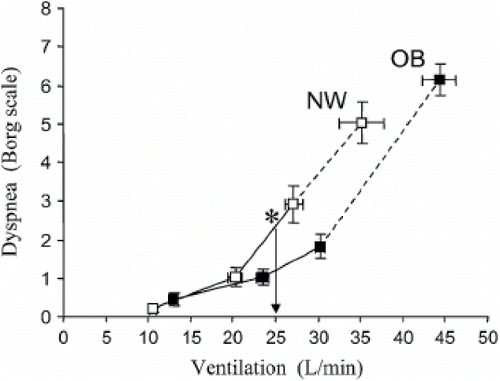
Figure 2. (A) Static lung volumes measured by body plethysmography at rest. Expiratory reserve volume (ERV) and functional residual capacity (FRC) (ERV + RV) were significantly (p < 0.05) lower in the obese (OB) group. (B) Lung volumes are shown from rest to peak exercise in OB patients COPD (closed squares) and in normal weight (NW) patients with COPD (open squares). In the OB compared with the NW group, end-expiratory lung volume (EELV) (standardized as a % of predicted TLC) was consistently lower (*p < 0.01) at rest and throughout exercise; the OB group reached an EELV at peak exercise that was similar to that of the NW group at the pre-exercise resting level. IC = inspiratory capacity; IRV = inspiratory reserve volume; VT = tidal volume (shaded area); RV = residual volume. Values are means ± SE. Reproduced from Ora J, Laveneziana P, Ofir D, Deesomchok A, Webb KA and O'Donnell DE. Combined effects of obesity and chronic obstructive pulmonary disease on dyspnea and exercise tolerance. Am J Respir Crit Care Med. 2009;180(10):964–71. Reprinted with permission of the American Thoracic Society. Copyright © 2017.
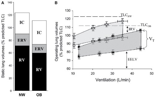
Figure 3. The adjusted predicted MRC score is given for each subject. For every given severity of airflow obstruction, obese patients are more dyspneic than normal weight patients. Regression lines for each BMI category also shown. (Normal weight: black dots; Overweight: gravy dots; Obese: white dots). Reproduced from Cecere L, Littman A. Obesity and COPD: associated symptoms, health-related quality of life and medication use. COPD 2011;8(4):275–284. Reprinted with permission of Taylor & Francis Ltd, http://www.tandfonline.com. Copyright © 2017.
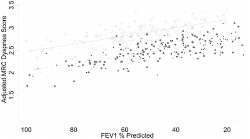
Figure 4. Post-bronchodilator lung volume components are shown divided by GOLD stage and BMI. UW = underweight; NW = normal weight; OW = overweight; OB = obese; RV = residual volume; ERV = expiratory reserve volume; IC = inspiratory capacity. Reproduced from O'Donnell DE, Deesomchok A, Lam YM, et al. Effects of BMI on static lung volumes in patients with airway obstruction. Chest. 2011;140 (Citation2):461–8. Reprinted with permission of Elsevier. Copyright © 2017.
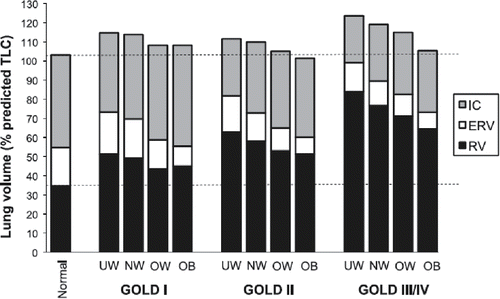
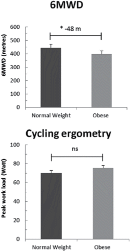
Figure 5. Main outcomes of cycle ergometry and 6-minute walk-test (6MWD) in normal weight and obese chronic obstructive pulmonary disease (COPD) patients. Reproduced from Maatman RC, Spruit MA, van Melick PP, et al. Effects of obesity on weight-bearing versus weight-supported exercise testing in patients with COPD. Respirology. 2016; 21(3):483–8. Reprinted with permission of John Wiley and Sons. Copyright © 2017.