Figures & data
Figure 1. IL-6 and IL-33 protein expression in bronchial mucosa samples, examples of rating based on the intensity of immunostaining (control – no staining; 1+, weak; 2+, intermediate; and 3+, strong).
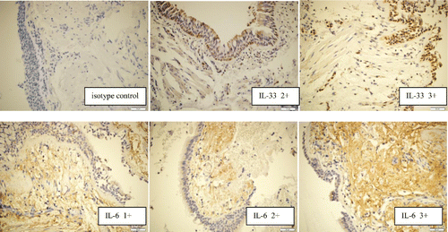
Table 1. Clinical characteristics of patients with asthma and COPD.
Table 2. Cytokine expression and cells count in different biological samples from patients with asthma and COPD.
Figure 2. Concentrations of IL-6 and IL-33 in serum, induced sputum (IS), and exhaled breath condensate (EBC) from patients with asthma and chronic obstructive pulmonary disease (COPD).
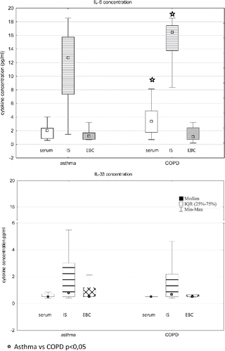
Table 3. Spearman correlation coefficients for relationships between the levels/expression of IL-6 and IL-33 in the investigated materials from patients with asthma.
Table 4. Spearman correlation coefficients for relationships between the levels/expression of IL-6 and IL-33 in the investigated materials from patients with chronic obstructive pulmonary disease (COPD).
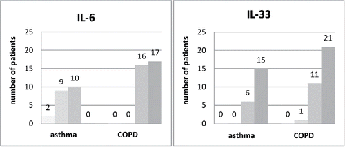
Figure 3. Distribution of tissue IL-6 and IL-33 protein expression in bronchial mucosa biopsy samples in patients with asthma (n = 21) and chronic obstructive pulmonary disease (n = 33). The X-axis presents the intensity of the immunostainings (■ 0, ■1+,■ 2+,■ 3+) separately for asthma and COPD groups.