
Figures & data
Table 1. The methylation levels of the perforin gene promoter in CD4 + T cells of the two Cell groups.
Figure 1. Histology of lung tissue. Notes: Lung tissue section was stained with HE. (A) Control rats receiving normal CD4 + T cells, adjuvant and PBS; (B) Rats receiving adjuvant and PBS; (C) Rats receiving CD4 + T cells hypomethylated, adjuvant and PBS. Data presented were one representative image data. Magnification: 400×. Abbreviations: HE, hematoxylin-eosin; PBS, phosphate buffered saline.
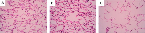
Figure 2. Expression of VEGF in rat lungs Notes: Expression of VEGF in rat lungs was detected by immunohistochemistry. (A) Control rats receiving normal CD4 + T cells, adjuvant and PBS; (B) Rats receiving adjuvant and PBS; (C) Rats receiving CD4 + T cells hypomethylated, adjuvant and PBS. Data presented were one representative image data. Magnification: 400×. Abbreviations: VEGF, Vascular endothelial growth factor; PBS, phosphate buffered saline.
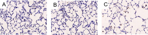
Table 2. The difference of MLI and MAN in the three groups.
Table 3. The difference of AECA in the three groups.
Table 4. The difference of VEGF OD in lung tissues in the three groups.
Figure 3. Apoptosis of alveolar septal cells in rat lungs. Notes: Apoptosis of alveolar septal cells in rat lungs was detected by immunohistochemistry. (A) Rats receiving CD4 + T cells hypomethylated, adjuvant and PBS; (B) Control rats receiving normal CD4 + T cells, adjuvant and PBS; (C) Rats receiving adjuvant and PBS. Data presented were one representative image data. Magnification: 400×. PBS, phosphate buffered saline.
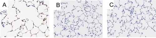
Table 5. The difference of AI of alveolar septal cells in the three groups.
Table 6. The methylation levels of the perforin gene promoter in CD4 + T cells in the three groups.