Figures & data
Figure 1 MAGE-D1 protein expression analyzed by Western blot. Ad-MAGE-D1 infection led to a marked over-expression of MAGE-D1 compared to mock- and Ad-GFP-infected HeLa cells.
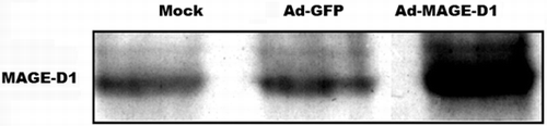
Figure 2 Ad-MAGE-D1 infection suppressed tumor cell proliferation. (A) and (B), Quantitative analysis of the proliferation of HeLa cells (A) and BEL7402 cells (B) infected with 50 MOI of the indicated adenovirus was performed by counting cells every 24 hours. Data are shown as cells/ml per well for triplicate wells from three independent experiments. *p < 0.05 vs. mock. (C) and (D), Cell proliferation for HeLa cells (C) and BEL7402 cells (D) was measured in a CCK-8 assay. Each dataset represents the ratio of the mock control after incubation for 24 h. Data are shown as the mean ± SD of three independent experiments.*p < 0.05 compared with the indicated mock control.
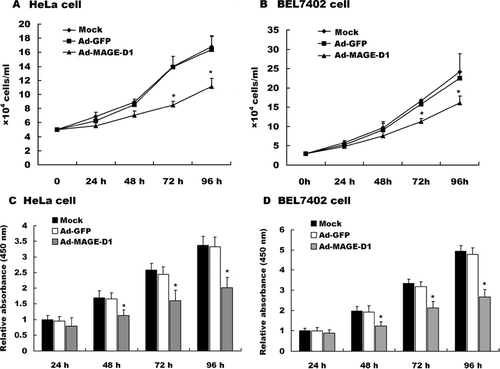
Figure 3 Ad-MAGE-D1 infection inhibited tumor cell migration in wound healing assay. (A) and (B), photomicrographs (× 200 magnification) showing the migration of HeLa cells infected with 50 MOI of the indicated adenovirus into the scraped area after 16 h under normoxic (A) or hypoxic (B) conditions, respectively. (C) and (D), Quantitative analysis of the migration for HeLa cells (C) and BEL7402 cells (D) after incubation for 16 h by counting the number of cells that had migrated to the wounded areas. Data are shown as the mean ± SD from three independent experiments. Results are expressed as a percentage of the migration in normoxic mock control. *p < 0.01 vs. mock.
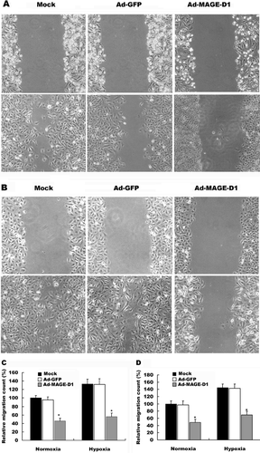
Figure 4 Ad-MAGE-D1 infection decreased the invasion of tumor cells. Cells transduced with 50 MOI of the indicated adenovirus for 48 h were plated on the transwell inserts, and transwell invasion assays were performed. Quantitative analysis of invasion after incubation for 8 h under normoxic or hypoxic conditions was performed by counting cells on membranes that were stained with Giemsa solution. Each experiment was performed in triplicate, and three separate experiments were performed. Data are expressed relative to normoxic mock condition. Results are shown as the mean ± SD *p < 0.01 vs. mock condition.
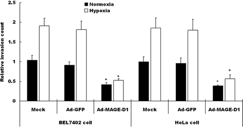
Figure 5 Ad-MAGE-D1 infection decreased adhesion of tumor cells to endothelial cells. (A) ECV304 cells were grown to confluence on glass cover slips in 6-well plates. A suspension of HeLa cells infected with the indicated adenovirus was added onto the monolayer of ECV304 cells. After 45 min of incubation at 37°C in normoxia (N) or hypoxia (H), cells were washed, counted, and photographed. (B) Quantitative evaluation of the data from adhesion of HeLa cells on the monolayer of ECV304 cells. Each assay was performed twice in triplicate. Results are shown as the mean ± SD, expressed as fold increase relative to normoxic mock control. *p < 0.01 vs. the indicated normoxic mock control.
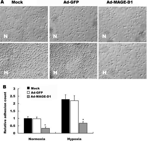
Figure 6 Ad-MAGE-D1 infection impaired lamellipodia formation and actin cytoskeletal reorganization induced by hypoxia in HeLa cells. Cells infected with 50 MOI of the indicated adenovirus were grown on glass cover slips in 6-well plates. After 4 h incubation in either normoxia (N) or hypoxia (H), cells were fixed and permeabilized, and f-actin structures were stained with rhodamine-conjugated phalloidin to visualize actin cytoskeleton organization (A), and the lamelipodia formation in migrating ECV304 cells (B). Scale bar, 10 μ m.
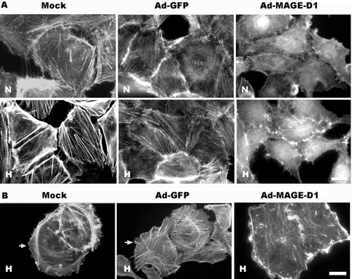
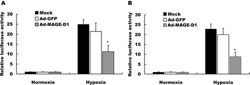
Figure 7 Ad-MAGE-D1 infection down-regulated hypoxia-induced HIF-1-dependent reporter gene expression. BEL7402 cells (A) and HeLa cells (B) infected with the indicated adenovirus were seeded in 6-well plates and transfected with 5xHRE/pGL3/VEGF/E1b, respectively. At 28 h post-transfection, cells were exposed to hypoxia for another 20 h. Luciferase and β -gal activities were measured. Relative luciferase activity was determined by the ratio of luciferase to β -gal activity and normalized to the value obtained in normoxic mock control cells. Data represent the mean ± SD of triplicate samples from a typical experiment, expressed as fold increase with respect to normoxic mock control. *p < 0.01 vs. the mean of hypoxic mock control cells.