Figures & data
Figure 1. Generating FAKloxP/loxPFN−/ − p53−/ − cell lines. (a) Genotyping of embryos from FAKloxP/loxPFN+/ − p53−/ − crosses, by PCR for FN. Embryos #1, #4, and #10 were wild-type, #5 and #8 were FN−/ − mutants, whereas others were FN+/ − . Genomic DNA isolated from primary wild-type MEF and FN−/ − cell line was used as a positive and negative control, respectively. (b) Outgrowth of fibroblast-like cells from E8.0–8.5 mouse embryo cultured in a drop of Matrigel for 3, 5, or 7 days. (c) Organization of FN matrix. Cells were cultured in the presence of 20 µg/ml exogenously added FN (Sigma) for 2 days. FN matrix organization is assessed by immunostaining of fixed and permeabilized cells. Rabbit polyclonal anti-FN antibody was from Sigma. FITC-conjugated donkey anti-rabbit antibody was from Jackson Immunoresearch.
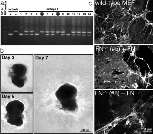
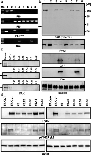
Figure 2. Generating FAK−/ − FN−/ − p53−/ − cell lines. (a) PCR verification of knocking out FAK in vitro. Marker lane (Ma): the 100-bp marker; lanes 1 and 2: FAK+/ + and FAK−/ − control cell lines, respectively; lane 3: parental FAKloxP/loxPFN−/ − p53−/ − line #5; lane 4: line #5 transduced with Cre-expressing adenovirus for 48 h; lane 5: single clone #5.41; lane 6: parental FAKloxP/loxPFN−/ − p53−/ − line #8; lane 7: line #8 transduced with Cre-expressing adenovirus for 48 h; lane #8: single clone #8.36. PCR product size: FN+, 900 bp; FN− 1060 bp; FAK+ 290 bp; FAKloxP, 400 bp; Cre, 419 bp. The same pair of primers is used to detect both FAK+ (290 bp) and FAK loxP (400 bp) alleles. (b) Western blot analyses of FAK knockout in vitro. Lane 1, FAK wild-type control cell line; lane 2, FAK-null control cell line; lane 3, cell line 5 (FAKloxP/loxPFN−/ − p53−/ − ); lane 4, cell line 5 (FAKloxP/loxPFN−/ − p53−/ − ) plus Cre; lane 5, cell line 5.41 (FAK−/ − FN−/ − p53−/ − ) 48 h after adding Cre; lane 6, cell line 8 (FAKloxP/loxPFN−/ − p53−/ − ); lane 7, cell line 8 (FAKloxP/loxPFN−/ − p53−/ − ) plus Cre; lane 8, cell line 8.36 (FAK−/ − FN−/ − p53−/ − ) 48 h after adding Cre. Arrow indicates size of FRNK, independently expressed C-terminal region of FAK. GFP and Cre expression are detected 48 h upon infection with adenovirus and they are gone in single cell clones. Paxillin used as a loading control. (c) Western blot analysis of FAK expression in clones derived from cell lines #5 and #8 after in vitro deletion of floxed region of FAK with adnovirus-delivered Cre. FAK+/ + , control cell line that express FAK; FAK−/ − , negative control line obtained directly from FAK−/ − embryos (Furuta et al. Citation1995). (d) Western blot analysis of Pyk2 expression and (auto)phosphorylation on Y402. FAK+/ + , control cell line that express FAK; FAK−/ − , negative control line obtained directly from FAK−/ − embryos (Ilic et al. Citation1995). Actin was used as a loading control. Anti-FAK antibodies were purchased from BD Transduction Laboratories and from Santa Cruz Biotechnology. Anti-Pyk2 antibody was from from BD Transduction Laboratories, anti-paxillin and ant-GFP from Zymed, anti-phosphoY402 Pyk2 from BioSource, anti-Cre from Covance. All secondary Abs were from Jackson Immunoresearch.