8,284
Views
50
CrossRef citations to date
0
Altmetric
Articles

Respiratory source control using a surgical mask: An in vitro study

, , &

Figures & data

Figure 1. Model of source manikin, receiver manikin, and environment interplay. Model of source manikin, receiver manikin, and environment interplay. Parameters can be set or measured. Dilution is an effect of the environment on the concentration of produced aerosols. Filtration (capture efficiency) is a function of the mask used and takes place at both the source and receiver. Particles that are not captured can be deflected (outward leakage around the faceseal perimeter) by the mask at the source and carried away from the receiver by the environmental flow. Breathing patterns simulate adults with tidal breathing or coughing.
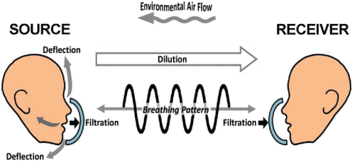
Figure 1. Model of source manikin, receiver manikin, and environment interplay. Model of source manikin, receiver manikin, and environment interplay. Parameters can be set or measured. Dilution is an effect of the environment on the concentration of produced aerosols. Filtration (capture efficiency) is a function of the mask used and takes place at both the source and receiver. Particles that are not captured can be deflected (outward leakage around the faceseal perimeter) by the mask at the source and carried away from the receiver by the environmental flow. Breathing patterns simulate adults with tidal breathing or coughing.

Figure 2. No flow chamber with, 0 air exchanges per hour (ACH). Schematic representation illustrating a chamber, containing the ventilated manikin heads 3 ft apart. Source head was connected to a nebulizer and exhaled radioactive aerosols. A filter was attached to the Receiver head to capture and quantify inhaled radioactive aerosols (exposure) (A). Hospital room chamber, with 6 ACH. Schematic representation illustrating a chamber, containing the ventilated manikin heads 3 ft apart. Source head was connected to a nebulizer and exhaled radioactive aerosols. A filter was attached to the Receiver head to capture and quantify inhaled radioactive aerosols (exposure) (B). Negative pressure room chamber, with 12 ACH. Schematic representation illustrating a chamber, containing the ventilated manikin heads 3 ft apart. Source head was connected to a nebulizer and exhaled radioactive aerosols. A filter was attached to the Receiver head to capture and quantify inhaled radioactive aerosols (exposure) (C).
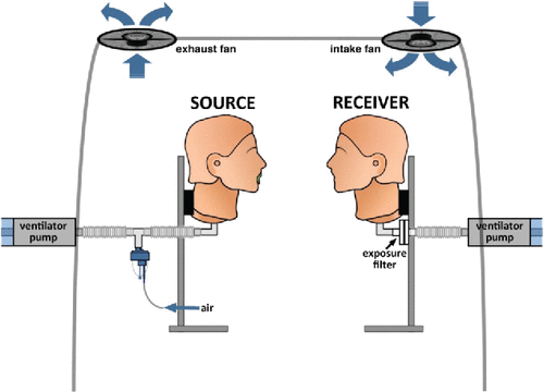
Figure 2. No flow chamber with, 0 air exchanges per hour (ACH). Schematic representation illustrating a chamber, containing the ventilated manikin heads 3 ft apart. Source head was connected to a nebulizer and exhaled radioactive aerosols. A filter was attached to the Receiver head to capture and quantify inhaled radioactive aerosols (exposure) (A). Hospital room chamber, with 6 ACH. Schematic representation illustrating a chamber, containing the ventilated manikin heads 3 ft apart. Source head was connected to a nebulizer and exhaled radioactive aerosols. A filter was attached to the Receiver head to capture and quantify inhaled radioactive aerosols (exposure) (B). Negative pressure room chamber, with 12 ACH. Schematic representation illustrating a chamber, containing the ventilated manikin heads 3 ft apart. Source head was connected to a nebulizer and exhaled radioactive aerosols. A filter was attached to the Receiver head to capture and quantify inhaled radioactive aerosols (exposure) (C).

Figure 3. Schematic representation illustrating cascade impaction experiments.

Figure 3. Schematic representation illustrating cascade impaction experiments.

Table 1. Particle distributions described by mass mean aerodynamic diameter (MMAD) and geometric standard deviation (GSD) for tidal breathing and cough. MMAD are expressed in μm.

Table 2. Respiratory protection factors (RPF) for tidal breathing and cough. An asterisk (*) denotes significance for a p-value <0.05 using the Kruskal-Wallis one-way analysis of variance.

Figure 4. Exposure data for tidal breathing, expressed as a percent of aerosol exhaled with a two-sided 95% CI, plotted for different masks on the Source or Receiver. An asterisk (*) denotes significance for a p-value <0.05 using the Kruskal-Wallis one-way analysis of variance. S = Source, R = Receiver, MaxEx = Maximum Exposure, SMnat = natural fit surgical mask, SF = SecureFit Ultra fitted surgical mask, N95 = 3M N95 respirator, N95vas = 3M N95 respirator with a Vaseline seal.
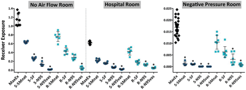
Figure 4. Exposure data for tidal breathing, expressed as a percent of aerosol exhaled with a two-sided 95% CI, plotted for different masks on the Source or Receiver. An asterisk (*) denotes significance for a p-value <0.05 using the Kruskal-Wallis one-way analysis of variance. S = Source, R = Receiver, MaxEx = Maximum Exposure, SMnat = natural fit surgical mask, SF = SecureFit Ultra fitted surgical mask, N95 = 3M N95 respirator, N95vas = 3M N95 respirator with a Vaseline seal.
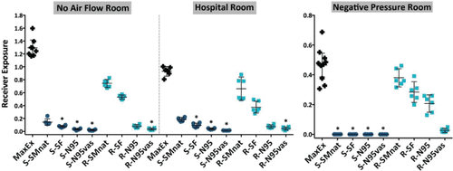
Figure 5. Exposure data for cough, expressed as a percent of aerosol exhaled with a two-sided 95% CI, plotted for different masks on the Source or Receiver. An asterisk (*) denotes significance for a p-value <0.05 using the Kruskal-Wallis one-way analysis of variance. S = Source, R = Receiver, MaxEx = Maximum Exposure, SMnat = natural fit surgical mask, SF = SecureFit Ultra fitted surgical mask, N95 = 3M N95 respirator, N95vas = 3M N95 respirator with a Vaseline seal.
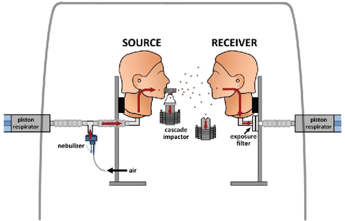
Figure 5. Exposure data for cough, expressed as a percent of aerosol exhaled with a two-sided 95% CI, plotted for different masks on the Source or Receiver. An asterisk (*) denotes significance for a p-value <0.05 using the Kruskal-Wallis one-way analysis of variance. S = Source, R = Receiver, MaxEx = Maximum Exposure, SMnat = natural fit surgical mask, SF = SecureFit Ultra fitted surgical mask, N95 = 3M N95 respirator, N95vas = 3M N95 respirator with a Vaseline seal.
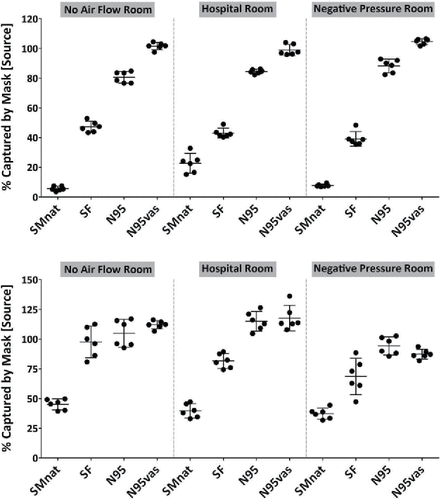
Figure 6. Mask capture efficiency (percent of aerosol exhaled with a two-sided 95% CI) mask on Source. (Upper Panel) Tidal Breathing. (Lower Panel) Cough.

Figure 6. Mask capture efficiency (percent of aerosol exhaled with a two-sided 95% CI) mask on Source. (Upper Panel) Tidal Breathing. (Lower Panel) Cough.