Figures & data
TABLE 1. Regenerative properties across vertebrate models of whole limb and digit tip regeneration after amputation.
FIGURE 1. Limb regeneration in axolotls. The axolotl follows a cellular mechanism of dedifferentiation and tissue-resident stem/progenitor cell activation for blastema formation (A) and subsequent differentiation to regenerate amputated limb (B).
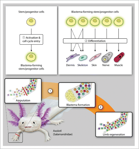
FIGURE 2. Limb regeneration in newts. Newt amphibians utilize a diverse array of cellular mechanisms for blastema formation (A), demonstrating both dedifferentiation and activation of both tissue-resident stem/progenitor cells and mature myocytes (muscle regeneration only). (While dedifferentiated dermal fibroblasts likely contribute to both dermal and skeletal regeneration, this has not been specifically shown in a newt model.) The cells forming the blastema then differentiate or re-differentiate to reconstitute the missing tissues of the amputated limb (B).
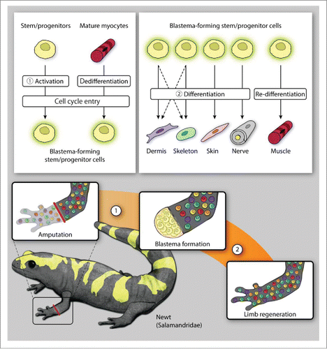
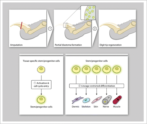
FIGURE 3. Digit tip regeneration in mammals. Mice employ cellular mechanisms that activate tissue-specific stem/progenitor cells for the formation of a blastema-like structure (A) which undergo germ-layer and lineage-restricted differentiation to reconstitute the distal digit tip (B).