1,235
Views
16
CrossRef citations to date
0
Altmetric
Research Paper

The effect of a silk Fibroin/Polyurethane blend patch on rat Vessels

, , , , &
Pages 115-124 | Received 02 May 2017, Accepted 14 Jun 2017, Published online: 13 Oct 2017

Figures & data

FIGURE 1. (A) ePTFE showed blood leakage from the suture site. (B) SF/Pellethane® swelled due to water absorption and blood did not leak.
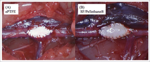
FIGURE 1. (A) ePTFE showed blood leakage from the suture site. (B) SF/Pellethane® swelled due to water absorption and blood did not leak.

FIGURE 2. (A) This figure was stained by MTC method. A 1-week sample from the control group revealed a clot between the ePTFE patch and its surrounding tissue. (B) After 3 weeks, inflammation cells and native tissue became non-reactive to ePTFE patches.
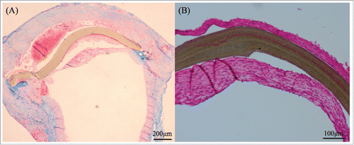
FIGURE 2. (A) This figure was stained by MTC method. A 1-week sample from the control group revealed a clot between the ePTFE patch and its surrounding tissue. (B) After 3 weeks, inflammation cells and native tissue became non-reactive to ePTFE patches.

FIGURE 3. This figure was shown samples of 3 months. SF/Pellethane® patch was stained by HE method (A) and shown collagen fibers in blue color by MTC staining (B). ePTFE patch was showed separation from the tissue (C) and was not infiltrated by collagen fibers (D).
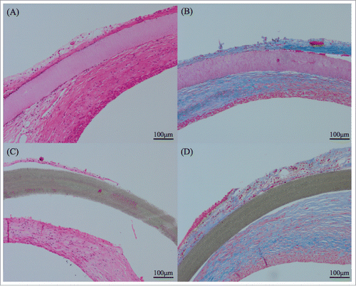
FIGURE 3. This figure was shown samples of 3 months. SF/Pellethane® patch was stained by HE method (A) and shown collagen fibers in blue color by MTC staining (B). ePTFE patch was showed separation from the tissue (C) and was not infiltrated by collagen fibers (D).

TABLE 1. Scoring of inflammation was done as follows; – (0), no; + (1), minute; ++ (2), modulate; +++ (3), severe and ± standard deviation (SD). Scoring of infiltration was done as follows; – (0), no; + (1), around patch; ++ (2), partial; +++ (3), overall and ± SD

TABLE 2. MTC staining results. Scoring of infiltration of collagen fibers was done as follows; – (0), no; + (1), a few or around patch; ++ (2), partial; +++ (3), overall and ± SD

TABLE 3. Scoring of calcification was done as follows; – (0), no; + (1), calcification deposits around patch; ++ (2), in patch; +++ (3), severe and ± SD

FIGURE 4. Kossa staining indicated calcification in brownish color. (A) ePTFE patch at 4 weeks had some calcified granules. (B) At 3 months, the calcification of the all ePTFE patch itself was observed. (C) SF/Pellethane® patches at a few samples had calcified granules. (D) At 3 months almost samples had only granules and did not react patch itself.
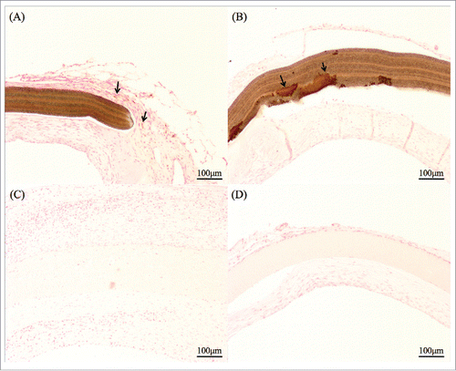
FIGURE 4. Kossa staining indicated calcification in brownish color. (A) ePTFE patch at 4 weeks had some calcified granules. (B) At 3 months, the calcification of the all ePTFE patch itself was observed. (C) SF/Pellethane® patches at a few samples had calcified granules. (D) At 3 months almost samples had only granules and did not react patch itself.

FIGURE 5. (A) Both patches were stained by CD31, and the stained region was only inside of the patch. The endothelial cells appeared along the vessel-like layer under the patch. (B) Also, the smooth muscle layer was formed along a native vessel and arranged clearly such as an arterial media.

FIGURE 5. (A) Both patches were stained by CD31, and the stained region was only inside of the patch. The endothelial cells appeared along the vessel-like layer under the patch. (B) Also, the smooth muscle layer was formed along a native vessel and arranged clearly such as an arterial media.

Reprints and Corporate Permissions

Please note: Selecting permissions does not provide access to the full text of the article, please see our help page How do I view content?

To request a reprint or corporate permissions for this article, please click on the relevant link below:

Academic Permissions

Please note: Selecting permissions does not provide access to the full text of the article, please see our help page How do I view content?

Obtain permissions instantly via Rightslink by clicking on the button below:

If you are unable to obtain permissions via Rightslink, please complete and submit this Permissions form. For more information, please visit our Permissions help page.