1,429
Views
0
CrossRef citations to date
0
Altmetric
Research Paper

Effects of porcine acellular dermal matrix treatment on wound healing and scar formation: Role of Jag1 expression in epidermal stem cells

, , , , , & show all
Pages 25-35 | Received 11 Dec 2017, Accepted 25 Jan 2018, Published online: 27 Apr 2018

Figures & data

Figure 1. Notch/Jagged1 signaling pathway was enhanced during wound healing. To investigate the role of Notch/Jagged1 signaling pathway in wound healing and scar formation in vivo, we conducted our experiments using a mouse skin wound model to assess the expression of several key factors of Notch/Jagged1 signaling pathway using western blot. N = 10 mice *P < 0.05 compared with day 0.
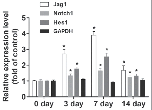
Figure 1. Notch/Jagged1 signaling pathway was enhanced during wound healing. To investigate the role of Notch/Jagged1 signaling pathway in wound healing and scar formation in vivo, we conducted our experiments using a mouse skin wound model to assess the expression of several key factors of Notch/Jagged1 signaling pathway using western blot. N = 10 mice *P < 0.05 compared with day 0.

Figure 2. Jagged 1 inhibited the differentiation of ESCs to MFB in vitro. (A) Representative immunofluorescence staining. a) Hoechst counterstaining, b) Keratin 15, c) β1 Integrin, and d) merged image. (B) Western blot analysis of α-SMA expression in ESCs.  (C) Western blot analysis of Collagen-I expression in ESCs. (D) Western blot analysis of Collagen-III expression in ESCs. *P < 0.05 compared with control.
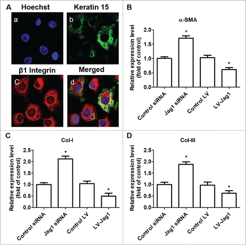
Figure 2. Jagged 1 inhibited the differentiation of ESCs to MFB in vitro. (A) Representative immunofluorescence staining. a) Hoechst counterstaining, b) Keratin 15, c) β1 Integrin, and d) merged image. (B) Western blot analysis of α-SMA expression in ESCs.  (C) Western blot analysis of Collagen-I expression in ESCs. (D) Western blot analysis of Collagen-III expression in ESCs. *P < 0.05 compared with control.

Figure 3. ESC-conditional knockout of Jag1 promoted scar formation. (A) Representative wound images, (B) Representative H&E staining, (C) Representative Masson staining. N = 10 mice/group.
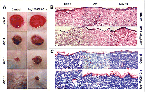
Figure 3. ESC-conditional knockout of Jag1 promoted scar formation. (A) Representative wound images, (B) Representative H&E staining, (C) Representative Masson staining. N = 10 mice/group.

Figure 4. Porcine ADM treatment accelerated wound closure and reduced scar formation in vivo. (A) α-SMA expression in wound tissue at day 7. (B) Collagen-I expression in wound tissue at day 7. (C) Collagen-III expression in wound tissue at day 7. (D) Representative wound images. Images were taken after carefully removed porcine ADM. (E) Wound area (%) over time. (F) Scar thickness. (G) Scar index. N = 10 mice/group. (A, B, C) *P < 0.05 compared with day 0, #P < 0.05 compared with VEH. (E, F, G) *P < 0.05 compared with control.
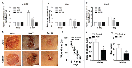
Figure 4. Porcine ADM treatment accelerated wound closure and reduced scar formation in vivo. (A) α-SMA expression in wound tissue at day 7. (B) Collagen-I expression in wound tissue at day 7. (C) Collagen-III expression in wound tissue at day 7. (D) Representative wound images. Images were taken after carefully removed porcine ADM. (E) Wound area (%) over time. (F) Scar thickness. (G) Scar index. N = 10 mice/group. (A, B, C) *P < 0.05 compared with day 0, #P < 0.05 compared with VEH. (E, F, G) *P < 0.05 compared with control.

Figure 5. Porcine ADM treatment increased Notch/Jag1 signaling. (A) Jag1 expression in wound tissue at day 7. (B) Notch1 expression in wound tissue at day 7. (C) Hes1 expression in wound tissue at day 7. N = 10 mice/group. *P < 0.05 compared with day 0, #P < 0.05 compared with VEH.

Figure 5. Porcine ADM treatment increased Notch/Jag1 signaling. (A) Jag1 expression in wound tissue at day 7. (B) Notch1 expression in wound tissue at day 7. (C) Hes1 expression in wound tissue at day 7. N = 10 mice/group. *P < 0.05 compared with day 0, #P < 0.05 compared with VEH.

Reprints and Corporate Permissions

Please note: Selecting permissions does not provide access to the full text of the article, please see our help page How do I view content?

To request a reprint or corporate permissions for this article, please click on the relevant link below:

Academic Permissions

Please note: Selecting permissions does not provide access to the full text of the article, please see our help page How do I view content?

Obtain permissions instantly via Rightslink by clicking on the button below:

If you are unable to obtain permissions via Rightslink, please complete and submit this Permissions form. For more information, please visit our Permissions help page.