Figures & data
Table 1. Summary of reported cases of labial minora fusion in the reproductive age
Table 2. Course of disease and clinical symptoms of 9 patients treated in our hospital
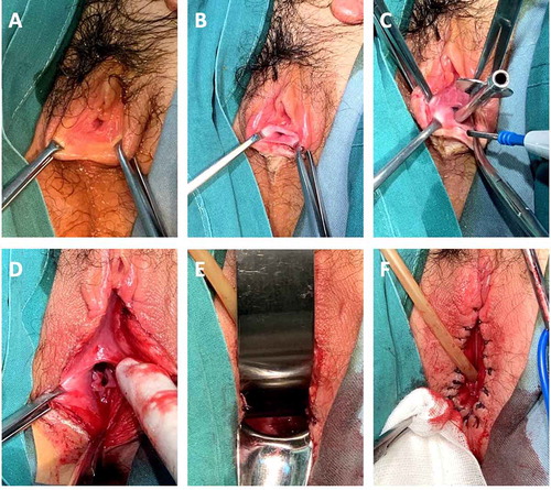
Figure 1. Procedure for labial fusion dissection. (a, b) Overview of the fused labial minora. Only the urethra meatus was visible. (c) A metal urethral catheter was inserted as a guide to avoid urethral injuries. (d) The fused labial minora were separated, and the vagina was revealed to have a normal width. (e) A normal cervix was evident. (f) The edges of the incision were sutured with 1/0 coated VICRYL separately