Figures & data
Figure 1. Isolated cells obtained from diseased explanted livers can be used to mimic the in vivo environment and become an effective experimental model to study a number of liver diseases including ALD, NAFLD, NASH, and metabolic diseases.
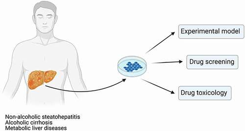
Table 1. Patient’s demographic data, yield and viability of isolated hepatocytes
Figure 2. (a) Example of NASH liver explant, (partial segments 2,3, Child-Pugh score C) before cell isolation; ongoing liver damage causing significant disruption of the normal hepatic architecture; surface of affected liver becomes irregular and nodular, with a progression of fibrosis. Representative phase-contrast images of cultured hepatocytes from NASH (b), MSUD (c), and ALD (d) livers, 24 hr culture. (e) Phase-contrast image of hepatocytes isolated from an ALD liver, recovered from cryopreservation, 24 h culture.
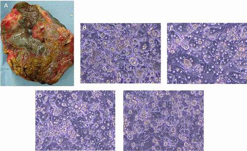
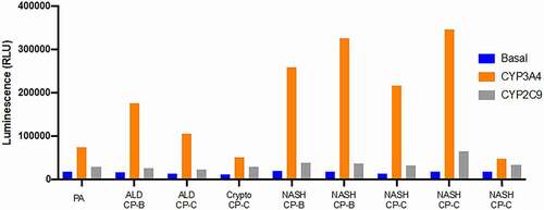
Figure 3. Detection of CYP3A4 and CYP2C9 gene induction in patient-derived hepatocytes isolated from 9 different diseased or cirrhotic explanted livers (PA, ALD, Crypto and NASH). CP: Child-Pugh.