Figures & data
Figure 1. Core molecules in the Hippo pathway and their mode of action.
As shown in the figure, the left half is the Hippo pathway in drosophila, and the right part is the Hippo pathway of mammal; the molecules at the corresponding position share the same or similar role.
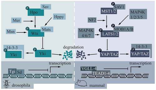
Figure 2. Hippo pathway controlling DPSCs.
Under a static magnetic field, both YAP and TAZ had a higher nuclear/cytosolic ratio and their downstream targets CTGF and ANKRD1 were found to be upregulated, while in the normal condition, YAP plays a predominant role in the nuclear transportation during odontogenic/osteogenic/neurogenic differentiation. Via the TGF-β/BMP pathway, TAZ was involved in regulating Cyr 61 and CTGF, hence associating proliferation and migration of DPSCs. Cytochalasin D could inhibit nuclei translocation of YAP/TAZ in both conditions.
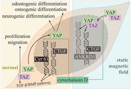
Figure 3. Hippo pathway controlling PDLSCs.
Functions of the Hippo pathway were illustrated separately. (3a) During proliferation of PDLSCs, more nucleus-translocated YAP contributed to upregulated expression of CDK6 and cyclin B1, which induced decreased G0/G1 and increased G2/M respectively. CDK6 inhibitors P18 and P27 inhibited. YAP-regulated cellular proliferation could be blocked by TNF-α. The ERK pathway was upregulated under overexpression of YAP. (3b) Overexpression of YAP could upregulate expression of OCN, COLI, RUNX2 and ALP activity for higher osteogenic potential and downregulate expression of PPARγ and C/EBPα for lower adipogenesis potential. Overexpression of YAP could also promote activity of β-catenin, hence forming crosstalk between Hippo and Wnt/β-catenin pathways. (3c) Postponed senescence was also seen under overexpression of YAP. For lower early and late apoptosis under overexpression of YAP, caspase and Bak, Bax, Bad, Bid and Bik of Bcl-2 family members were found to be inhibited.
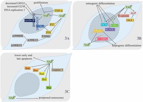
Figure 4. Development of enamel organ.
Formation of enamel starts from interaction of ectoderm and mesenchyme. Thickened dental lamina could form dental placode, and then it comes to the bud stage. During the subsequent cap stage, enamel organ is formed, where enamel knot was seen. Continuous growth of the cap stage will transfer into the bell stage, and differentiated ameloblasts and odontoblasts contributed to formation of enamel and dentin.
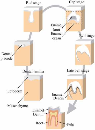
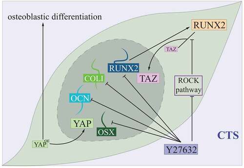
Figure 5. Hippo controlling PDLCs under the cyclic tensile stress condition.
Under CTS, both YAP and TAZ had a higher nuclear/cytosolic ratio. RUNX2, one of the osteogenic proteins, showed higher interaction of TAZ, while this effect could be reversed by ROCK pathway inhibitor Y27632. With Y27632, osteogenic mRNA involving COLI, OSX, OCN and RUNX2 weas seen to be inhibited. Overexpression of YAP was related to promoted osteoblastic differentiation under CTS.