
Figures & data
Figure 1. Attenuation of cardiac injury by exosomes from TNF-α-treated BMSCs in the MI mouse model.
(a) Study design of animal experiments to investigate the impact of exosomes derived from TNF-α-treated BMSCs on cardiac injury post-MI. At 28 days post-MI, serum levels of key cardiac injury markers, including LDH (Lactate Dehydrogenase) (b), CK-MB (Creatine Kinase-MB) (c), and Troponin I (d), were quantified. These analyses served as pivotal indicators in assessing the therapeutic efficacy of the administered exosomes. Data were shown with mean ± SD. 10 mice were used for each group. *p < .05, **p < .01, ***p < .001 from Brown-Forsythe ANOVA test followed by Dunnett’s T3 multiple comparisons test.
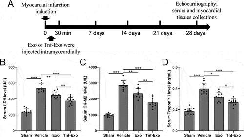
Figure 2. Preservation of cardiac function by exosomes from TNF-α-treated BMSCs in the MI mouse model.
The protective effects of exosomes derived from TNF-α-treated BMSCs on cardiac function post-MI is evidenced through comprehensive assessments of key parameters, including Left Ventricular Ejection Fraction (LVEF) (a), Left Ventricular Fractional Shortening (LVFS) (b), Left Ventricular Internal Diameter at end-diastole (LVIDd) (c), and Left Ventricular Internal Diameter at end-systole (LVIDs) (d) at the 28-day mark post-MI. These comparisons elucidate the therapeutic impact of the administered exosomes on preserving overall cardiac function. Data were shown with mean ± SD. 10 mice were used for each group. *p < .05, **p < .01, ***p < .001 from Brown-Forsythe ANOVA test followed by Dunnett’s T3 multiple comparisons test.
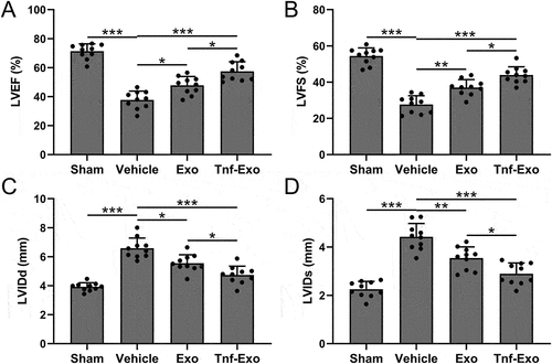
Figure 3. Exosomes derived from TNF-α-treated BMSCs alleviated infarct size after myocardial infarction in mice.
(a) The calculations of left ventricular wall thickness and (b) infarct ratio in the experimental groups. Data were shown with mean ± SD. 6 mice were used for each group. *p < .05, **p < .01, ***p < .001 from Brown-Forsythe ANOVA test followed by Dunnett’s T3 multiple comparisons test.
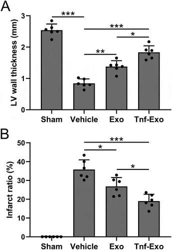
Figure 4. Exosomes derived from TNF-α-treated BMSCs enhanced M2 macrophage polarization in the myocardial tissues after myocardial infarction in mice.
The percentage of M1 (a) and M2 (b) polarized macrophages in the total number of macrophages in the infarct area of myocardial tissues at 28 days post-MI. Data were shown with mean ± SD. 6 mice were used for each group. *p < .05, **p < .01, ***p < .001 from Brown-Forsythe ANOVA test followed by Dunnett’s T3 multiple comparisons test.
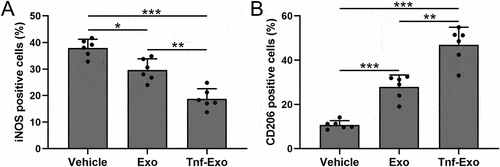
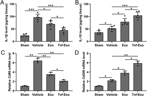
Figure 5. Exosomes derived from TNF-α-treated BMSCs enhanced M2 macrophage polarization in the myocardial tissues after myocardial infarction in mice.
ELISA was used to measure the levels of IL-1β (a) and IL-10 (b) in left ventricular tissues at 28 days post-MI. 6 mice were used for each group. qRT-PCR was used to measure the mRNA expressions of Cd86 (c) and Cd206 (d) in left ventricular tissues at 28 days post-MI. 6 mice were used for each group and the experiments were repeated 3 times using mixed tissue homogenates. Data were shown with mean ± SD. *p < .05, **p < .01, ***p < .001 from Brown-Forsythe ANOVA test followed by Dunnett’s T3 multiple comparisons test.
Data availability statement
The raw data supporting the conclusions of this article will be made available by the authors, without undue reservation.