
Figures & data
Figure 1. The simplest types of knots. The simplest mathematical knots are shown in panel (a) and are labeled by the number of crossings in the simplest two-dimensional projection followed by a conventional indexing subscript. The corresponding physical knots, which are obtained by opening the closed chains, are shown in panel (b).
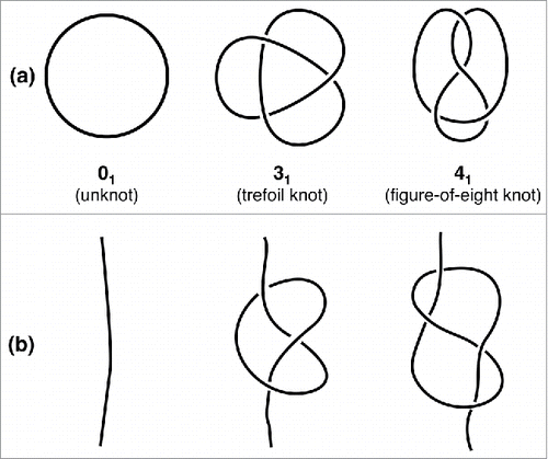
Figure 2. RNA chain length and types. The distribution of lengths (number of phosphates) of all 7,013 RNA molecules with more than five phosphates deposited in the PDB as of August 2015 is shown. The color code describes the contribution of each categorized subdivision to the total amount of chain.
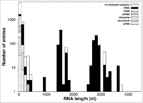
Table 1. Knotted RNAs in the survey data set. The knots are likely artifactual results of cryo-em reconstruction. The entries are the same as those shown in of ref. 20, though some of the PDB codes differ because the PDB introduced a new archiving system of large structures (with no change of atomic coordinates). The overall knot type was established by using the minimally-invasive closing procedure of ref. 46. The # sign in the knot label of the last entry denotes the composition (concatenation) of various prime knots.
Figure 3. Possible pathways leading to RNA knotting. The sketch illustrates possible pathways through which two sets of interleaved self-complementary sequences (marked with squares and circles, respectively) can fold and produce a knotted RNA structure. The latter is tied in a figure-of-eight knot and the diagram representing the two helices features the characteristic pseudoknot crossings.
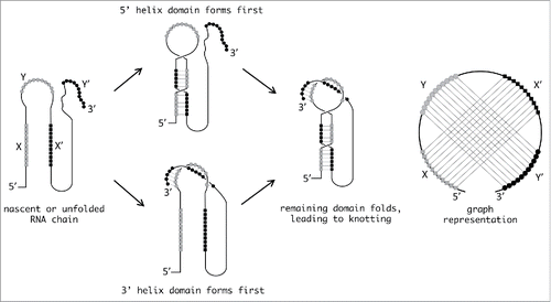
Table 2. Candidate knot-forming RNA molecules from Pseudoknot database (http://ekevanbatenburg.nl/PKBASE/PKBGETSUM.HTML#TOP; updated November 28, 2014, accessed October 11, 2015).