
Figures & data
Figure 1. CTIF inhibits HIV-1 and HIV-2 Gag synthesis
A) Jurkat cells were infected with VSV G-pseudotyped HIV-1 and cell extracts prepared at 0, 8, 12, 20, 24, 48, 72 and 96 hours post-infection were used to detect Gag (and its processing intermediates) and CTIF by Western blot. GAPDH was used as loading control. Band intensity for CTIF and Gag (pr55) from three independent experiments were quantified, normalized to GAPDH and plotted (n = 3, ± SEM). B) SCR-KD or CTIF-KD cells (1x10⁵ cells/well) were transfected with 1 µg of pcDNA-d2EGFP (used as a control) or pcDNA3-Flag-CTIF together with 0.3 µg of pNL4.3R as described in materials and methods. Gag-Renilla activity was determined at 24 hpt. Results were normalized to the control (arbitrary set to 100%) and correspond to the mean ± SD of three independent experiments (*P < 0.05; **P < 0.01 and ****P < 0.0001, t-test). Cells extracts were used to verify Flag-CTIF and d2EGFP expression by Western blot. GAPDH was used as a loading control.C) Jurkat cells were infected with the VSV G-pseudotyped HIV-1 produced under control and Flag-CTIF expression and cellular extracts prepared at 48 hours post-infection were used to detect Gag and its processing intermediates. GAPDH was used as loading control. This image corresponds to a representative Western blot from three independent experiments.D) SCR-KD or CTIF-KD cells (1x10⁵ cells/well) were transfected with 0.3 µg of the HIV-2 pROD10R reporter proviral DNA as described in materials and methods. Gag-Renilla activity was determined at 24 hpt. Results were normalized to the control (arbitrary set to 100%) and correspond to the mean ± SD of three independent experiments (*P < 0.05, t-test).E) HeLa cells (1x10⁵ cells/well) were transfected with 1 µg of pcDNA-d2EGFP (used as control) or pcDNA3-Flag-CTIF together with 0.3 µg of pROD10R as described in materials and methods and Gag-Renilla activity was determined at 24 hpt. Results were normalized to the control (arbitrary set to 100%) and correspond to the mean ± SD of three independent experiments (***P < 0.001; t-test). In parallel, cells extracts were used to detect Flag-CTIF by Western blot. GAPDH was used as a loading control.
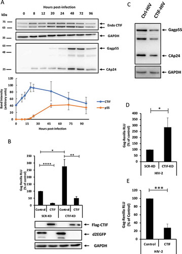
Figure 2. CTIF acts on Rev function
A) HeLa cells (8x10⁴ cells/well) transfected with 1 µg pEGFP-Rev were subjected to the proximity ligation assay using a rabbit anti-CTIF antibody diluted 1/100 and a mouse anti-GFP antibody diluted 1/100 together with the Duolink® in situ kit as described in materials and methods. Mock corresponds to non-transfected cells. The PLA dots are shown in red and DAPI in blue. Scale bar 10 µm. Quantification of dots per cell in Mock-CTIF (n = 34 cells) and Rev-CTIF (n = 33 cells) are presented (****P < 0.0001, Mann–Whitney test).B) SCR-KD and CTIF-K cells (1x10⁵ cells/well) were transfected with 0.3 µg of pNL4.3R or pNL4.3R-ΔRev as described in materials and methods and Renilla luciferase activity was determined at 24 hpt. Results were normalized to the control (arbitrary set to 100%) and correspond to the mean ± SD of three independent experiments (*P < 0.05, t-test).C) SCR-KD or CTIF-KD cells(1x10⁵ cells/well) were transfected with 0.3 µg of pNL4.3 R as described in materials and methods. Gag-Renilla activity (left panel) and cytoplasmic full-length RNA levels (right panel) were determined at 24 hpt. Results were normalized to the control (arbitrary set to 100%) and correspond to the mean ± SD of three independent experiments (***P < 0.001, *P < 0.05, t-test).D) HeLa cells (1x10⁵ cells/well) were transfected with 1 µg of pcDNA-d2EGFP(used as a control) or pcDNA-Flag-CTIF together with 0.3 µg pNL4.3 R (left panel) or pNL4.3 R-CMV-ΔU3/U5 (right panel) as described in materials and methods. Results were normalized to the control (arbitrary set to 100%) and correspond to the mean ± SD of three independent experiments (**P < 0.01, ****P < 0.0001, t-test t-test).E) Conservation analysis of the AGGA loop (nucleotides 234 to 247 in NL4.3) from the 5ʹ-UTR of 879 HIV-1 isolates.
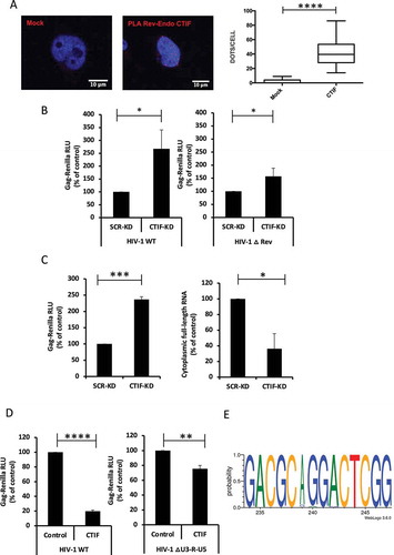
Figure 3. CTIF inhibits Gag synthesis through its N-terminal domain
A) Upper panel: Scheme of CTIF showing its N-terminal CBP80-binding domain and its C-terminal MIF4G domain. Numbers indicate amino acid positions. Bottom panel: HeLa cells (1x10⁵ cells/well) were transfected with 1 µg of pcDNA-d2EGFP (used as a control), pcDNA3-Flag-CTIF, pcDNA3-Flag-CTIF-N-ter or pcDNA-Flag-CTIF-C-ter together with 0.3 µg of pNL4.3R as described in materials and methods. Gag-Renilla activity was determined at 24 hpt. Results were normalized to the control (arbitrary set to 100%) and correspond to the mean ± SD of three independent experiments (**P < 0.01; NS = non-significant, t-test). Cells extracts were used to detect Flag-CTIF by Western blot. Actin was used as a loading control.B) HeLa cells (8x10⁴ cells/well) transfected with 1 µg of pEGFP-Rev together with 1 µg of pcDNA3-Flag-CTIF, pcDNA3-Flag-CTIF-N-ter or pcDNA-Flag-CTIF-C-ter were subjected to the proximity ligation assay using a rabbit anti-Flag antibody diluted 1/200 and a mouse anti-GFP antibody diluted 1/100 together with the DuolinkⓇ in situ kit as described in materials and methods. Mock corresponds to non-transfected cells. The PLA signal is shown in red and DAPI in blue. Scale bar 10 µm. Quantification of dots per cell in Rev-CTIF (n = 29 cells), Rev-CTIF-N-ter (n = 43 cells) and Rev-CTIF-C-ter (n = 43 cells) are presented (****P < 0.0001, NS; non-significant, Mann–Whitney test).C) HeLa cells (6x10⁶ cells/plate) were transfected with 10 µg of pNL4.3 together with 10 µg pcDNA3-Flag-CTIF and 3 µg of pEGFP or pEGFP-Rev as indicated. At 24 hpt, cell extracts were used for RNA immunoprecipitation as described in materials and methods. Results are expressed as the IP/input ratio normalized to the pNL4.3 condition (arbitrary set at 100%). Results correspond to the average ± SEM of three independent experiments. (***P < 0.001; NS = non-significant, t-test). Cells extracts were used to detect Flag-CTIF, EGFP and EGFP-Rev by Western blot.D) HeLa cells (8x10⁴ cells/well) transfected with 0.3 µg of pNL4.3 together with 1 µg pcDNA3-Flag-CTIF, pcDNA3-Flag-CTIF-N-ter or pcDNA-Flag-CTIF-C-ter were subjected to ISH-PLA using a rabbit anti-Flag antibody diluted 1/200 and a mouse anti-digoxigenin antibody diluted 1/100 together with the DuolinkⓇ in situ kit as described in materials and methods. Mock corresponds to non-transfected cells. Quantification of dots per cell in full-length RNA-CTIF (n = 36 cells), full-length RNA-CTIF-N-ter (n = 30 cells) and full-length RNA-CTIF-C-ter (n = 27 cells) are presented (****P < 0.0001, NS; non-significant, Mann–Whitney test).
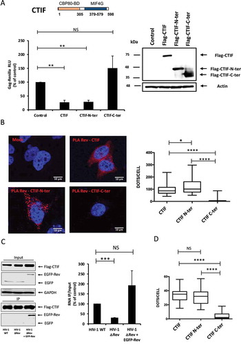
Figure 4. CTIF modifies Rev localization within the cell
A) HeLa cells (8x10⁴ cells/well) transfected with 1 µg pEGFP-Rev together with 1 µg of pcDNA3-Flag-CTIF, pcDNA3-Flag-CTIF-N-ter, pcDNA3-Flag-CTIF-C-ter or pcDNA-β-globin (used as a control) were subjected to immunofluorescence assay using a rabbit anti-Flag antibody. Phase contrast signal is shown in grey, EGFP-Rev signal in green, Flag-tag signal in magenta and DAPI in blue. Scale bar 10 µm.B) Quantification of the sub-cellular localization of EGFP-Rev in conditions presented in A) (*P < 0.05; NS; non-significant, t-test).C) HeLa cells (8x10⁴ cells/well) transfected with 1 µg of pEGFP-Rev together with 1 µg of pcDNA3-Flag-CTIF, pcDNA3-Flag-CTIF-N-ter or pcDNA3-Flag-CTIF-C-ter were subjected to the proximity ligation assay using a rabbit anti-Flag antibody and a mouse anti-GFP antibody and the DuolinkⓇ in situ kit. After the rolling circle amplification of the proximity ligation assay, cells were incubated with Alexa anti-rabbit 647 secondary antibody as described in materials and methods. Mock corresponds to non-transfected cells. The PLA signal is shown in red, EGFP-Rev signal in green, Flag signal in magenta and DAPI in blue. Scale bar 10 µm.
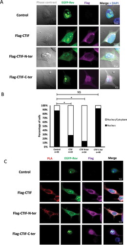
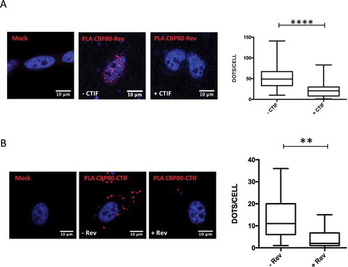
Figure 5. And interferes with the Rev-CBP80 interaction
A) HeLa cells (8x10⁴ cells/well) transfected with pEGFP-Rev and pCMV-myc-CBP80 together with pcDNA β-globin (- CTIF condition) or pcDNA3-Flag-CTIF (+ CTIF condition) were analyzed for the Rev-CBP80 interaction by PLA using a rabbit anti-GFP antibody diluted 1/100 and a mouse anti-Myc antibody (Sigma-Aldrich) diluted 1/100 together with and the DuolinkⓇ in situ kit as described in materials and methods. Mock corresponds to non-transfected cells. The PLA signal is shown in red and DAPI in blue. Scale bar 10 µm. Quantification of dots per cell in – CTIF (n = 43 cells) and + CTIF (n = 37 cells) is presented (****: P < 0.0001, Mann–Whitney test).B) HeLa cells (8x10⁴ cells/well) transfected with pcDNA3-Flag-CTIF and pcDNA-V5-CBP80 together with pEGFP (- Rev condition) or pEGFP-Rev (+ Rev condition) were analyzed for the CTIF-CBP80 interaction by PLA using a rabbit anti-Flag antibody diluted 1/200 and a mouse anti-V5 antibody (Santa Cruz) diluted 1/100 together with the DuolinkⓇ in situ kit as described in materials and methods. Mock corresponds to non-transfected cells. The PLA signal is shown in red and DAPI in blue. Scale bar 10 µm. Quantification of dots per cell in – Rev (n = 46 cells) and + Rev (n = 40 cells) is presented (**: P < 0.01, Mann–Whitney test).
Supplemental material