Figures & data
Figure 1. Single molecule methods of mRNA detection
A – Single molecule FISH (smFISH). 10 probes with 5 fluorophores each are used for mRNA detection with single molecule sensitivity [Citation8]. B – Single molecule inexpensive FISH (smiFISH). The mRNA is detected using non-labelled primary probes, which contain a sequence complementary to target RNA and a read-out sequence identical for all the probes. This read-out sequence hybridizes to a unique secondary probe labelled with 2 fluorophores on its 3ʹ and 5ʹ [Citation13]. C – Sequential smFISH, example of Multiplexed Error Robust FISH (merFISH). RNA targets are first hybridized with encoding probes, containing sequence complementary to target and two read-out sequences. Each RNA target is identified by a combination of uniques read-out sequences in subsequent rounds of hybridization with secondary probes, one secondary probe per round of hybridization. After each round of hybridization with a secondary probe, the signal is detected, registered and the probe is removed. Each RNA is identified with a unique barcode (lower panel, left column), in which a hybridization round functions as a bit. If the RNA gives a signal with a given probe it is assigned 1, in case of no signal it is assigned 0. To increase the sensitivity, sets of 24–100 primary probes are used per mRNA (top panel, left) [Citation16].
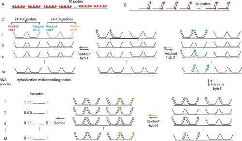
Figure 2. Different patterns of mRNA localization revealed by FISH and smFISH
A – FISH on Drosophila embryo (mRNA green/nuclei red). The gene names are indicated on the bottom left and localization patterns above the photos. From left to right: bicoid mRNA displays anterior localization in the embryo; anillin mRNA has perinuclear localization; CG1962 mRNA localizes to microtubules networks and centrosomes during cell division. Reprinted from Lécuyer E, Yoshida H, Parthasarathy N, Alm C, Babak T, Cerovina T, Hughes TR, Tomancak P, Krause HM. Global analysis of mRNA localization reveals a prominent role in organizing cellular architecture and function. Cell 2007; 131:174–87. Pages 178, 180, 181. Copyright 2020, with permission from Elsevier. B – smFISH on Hela cells. The genes names are indicated on the bottom left and localization patterns are above the photos. Top – from left to right: KIF5B is localized to the cell protrusions; FLNA is localized to the cell edge; BUB1 – cytoplasmic foci. Orange arrows indicate mentioned locations. Bottom – ASPM mRNA is localized to nuclear envelope and cytoplasmic foci in interphase and to spindle pole during cell division. Left – smFISH, right – nuclei stained with DAPI. Orange arrow indicates interphase cell; pink arrow – mitotic cell. Reprinted from Chouaib R, Safieddine A, Pichon X, Imbert A, Kwon OS, Samacoits A, Traboulsi A-M, Robert M-C, Tsanov N, Coleno E, et al. A Dual Protein-mRNA Localization Screen Reveals Compartmentalized Translation and Widespread Co-translational RNA Targeting. Dev Cell 2020; 54:773–791.e5. Pages 775, 776. Copyright 2020, with permission from Elsevier.
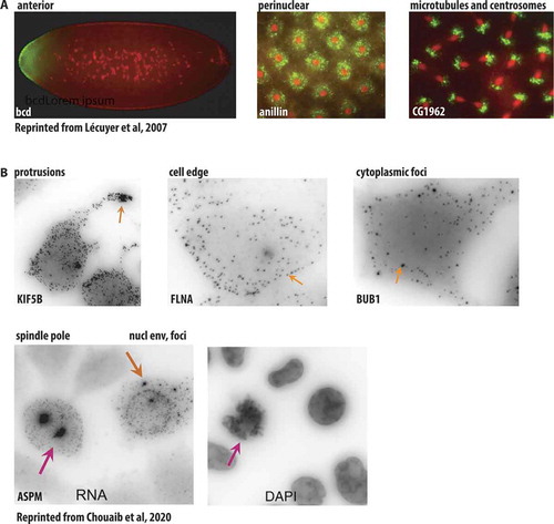
Figure 3. RNA detection in living cells
A – Molecular beacons. Unbound MBs are not fluorescent due to a proximity of a quencher (blue circle) to a fluorophore (red circle). Upon binding to the target RNA, these interactions are disrupted leading to a fluorescence emission. B – Principle of fluorogenic RNA aptamers. RNA aptamers are based on the interaction of a specific RNA structure with a fluorogenic dye (green circle), which becomes fluorescent upon aptamer binding. C – The regulatory RNA aptamer Pepper. Pepper RNA binds a protein destabilization domain fused to a fluorescent protein, leading to its stabilization and allowing the fluorescent signal to be observed [Citation54]. D – MCP-GFP detection of mRNA in live cells. MCP-GFP binds as a dimer to stem-loops from bacteriophage MS2. These stem-loops are usually inserted in 3ʹUTR of the mRNA of interest, and 24 stem-loops enable detection of single-RNA molecules [Citation55,Citation56].
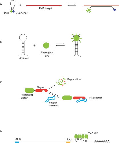
Figure 4. mRNP transport complexes
A – ASH1 mRNA in yeast Saccharomyces cerevisiae. ASH1 is transported on F-actin cables, it is bound by RBP She2p co-transcriptionally, an adaptor She3p links RNA-She2p complex to a motor Myo4D in the cytoplasm [Citation126]. B – RNA-stimulated assembly of transport complex for localized RNAs in Drosophila embryo. RBP Egalitarian (Egl) binds the RNA LE and promotes its interaction with the dimer of Bicaudal-D (BicD). RNA:Egl complex binding to BicD releases its autoinhibitory loop facilitating the interaction with dynein to form an active dynein-dynactin complex for the transport to the minus end of microtubules. Two Egl proteins are associated with one molecule of RNA [Citation144]. C – Transport of septin mRNAs in fungus Ustilago maydis. mRNAs are bound by the RBP Rrm4 together with its partner Grp1 and transported on Rab5a positive early endosomes. The complex mRNA-Rrm4-Grp1, containing also Poly-A binding protein 1 (Pab 1), is tethered on endosomes through the adaptor Upa1. The mRNA is translated during the transport [Citation184]. D – Model of β-catenin mRNA translational regulation. β-catenin mRNA accumulates in cytoplasmic translation factories, where it is translated and the newly-made β-catenin protein is degraded by the destruction complex. Upon WNT signalling activation, the factories are dissolved and the newly synthesized β-catenin migrates to the nucleus to activate transcription [Citation31,Citation192].
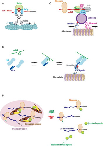
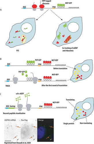
Figure 5. Single-molecule approaches to study mRNA translation in live cells
A – Tracking of mRNAs and ribosomes. RNAs are labelled with MCP-GFP (top, green ovals) and ribosomes with red fluorescent protein (top, red ovals), allowing to follow them by Fluorescence Fluctuation Spectroscopy (FFS) or single-particle tracking. Left: two-photon FFS monitors passages of mRNAs and ribosomes through a microscopic volume in the cell [Citation146]. Right: single particle co-tracking of mRNPs and ribosomes [Citation147]. B – TRICK. The mRNA is labelled with PCP-GFP (left, green ovals) in its coding region and with MCP-RFP (left, red ovals) in its 3ʹ-UTR. Ribosome removes bound PCP-GFP during translation. The non-translated mRNAs are visible in the microscope as yellow dots due to the superposition of green and red colours (right), while after the first round of translation the mRNAs become red dots (right) [Citation88]. C – Visualization of nascent translation. Top left – The mRNA is labelled with MCP-RFP (left, red ovals), the nascent peptide is visualized using single chain antibodies fused to super-folder GFP ScFv-sfGFP (left, green stars), that recognize SunTag, or with anti-Flag antibodies, or Frankenbody in case of protein labelling with repeats of Flag- or HA- tags. Right – Non-translating mRNAs are visible as red dots (right), the single molecules of protein are green dots (right) and actively transcribed mRNA are yellow dots [Citation149]. Bottom – Micrograph of HeLa cells with a SunTagged ASPM allele, showing ASPM mRNA (by smiFISH, left and red), the signal from the SunTag (middle and green); blue, nuclear staining with DAPI. White and black arrows, a single mRNA positive for the SunTag; orange arrow, an mRNA foci positive for the SunTag. Scale bar: 10 mm. Reprinted from Chouaib R, Safieddine A, Pichon X, Imbert A, Kwon OS, Samacoits A, Traboulsi A-M, Robert M-C, Tsanov N, Coleno E, et al. A Dual Protein-mRNA Localization Screen Reveals Compartmentalized Translation and Widespread Co-translational RNA Targeting. Dev Cell 2020; 54:773–791.e5. Page 781, Copyright 2020, with permission from Elsevier.