
Figures & data
Figure 1. Workflow diagrams of PBL and CBRPP
(A) Workflow diagram of PBL. By fusing protein of interest to enzyme that generate reactive molecules, most commonly biotin, adjacent proteins are covalently labelled so that they can be isolated by streptavidin beads and identified by mass spectrometry.(B) Workflow diagram of CBRPP. By fusing dCas13 and PBL enzyme together, dCas13, under the guidance of a specific crRNA, acts as an RNA tracker to bring PBL enzyme to the target RNA, then PBL enzyme biotinylates surrounding proteins of the target RNA, followed by streptavidin beads enrichment of biotinylated proteins and mass spectrometry.(C) Workflow of transient transfection of dPspCas13b-APEX2-NES and Psp-crRNAs to identify the RBPs of ACTB mRNA. Psp-NT1-2: non-targeting Psp-crRNA 1 and 2. ACTB-Psp-crRNA3-4: ACTB Psp-crRNA 3 and 4.
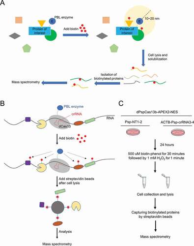
Table 1. crRNAs used in this paper
Table 2. qPCR primers used in this paper
Figure 2. dRfxCas13d is not suitable for CBRPP to study RNA-protein interactions
(A) Plasmids used in this figure. NLS: nuclear localization sequence. NES: nuclear export sequence. EGFP: enhanced green fluorescent protein. T2A: T2A self-cleaving peptide. HA: haemagglutinin tag.(B) Upper: HEK293T cells were co-transfected with RfxCas13d/RfxCas13d-APEX2/APEX2-RfxCas13d and B4GALNT1 Rfx-crRNA to detect the mRNA level of B4GALNT1 by RT-qPCR after 48 hours. Rfx-NT1: non-targeting Rfx-crRNA 1; Rfx-NT2: non-targeting Rfx-crRNA 2. B4-Rfx-crRNA: B4GALNT1 Rfx-crRNA. Bottom: western blot to measure the protein expression level of RfxCas13d, APEX2-RfxCas13d and RfxCas13d-APEX2.(C) HEK293T cells were co-transfected with RfxCas13d and ACTB Rfx-crRNAs to detect the mRNA level of ACTB by RT-qPCR after 48 hours.(D) Representative images for dRfxCas13d-APEX2-NES imaging with two ACTB Rfx-crRNAs targeting ACTB mRNA in HEK293T. Mock: no treatment. Sodium arsenite: treating cells with 0.5 mM sodium arsenite for 30 minutes. Stress granules are indicated by G3BP1 staining. Scale bars, 10 μm.For B and C: n = 3, mean ± SD, two-sided Student’s t-test, ****P < 0.0001; ***P < 0.001; **P < 0.01; *P < 0.05; ns: not significant.
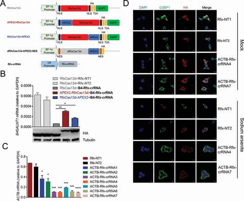
Figure 3. Transient transfection of dPspCas13b-APEX2-NES to identify the RBPs of ACTB mRNA
(A) Plasmids used in this figure. NES: nuclear export sequence. EGFP: enhanced green fluorescent protein. P2A: P2A self-cleaving peptide. HA: haemagglutinin tag. Linker: 3x(GGGGS), G: glycine, S: serine.(B) HEK293T cells were co-transfected with PspCas13b/PspCas13b-APEX2 and ACTB Psp-crRNAs to detect the mRNA level of ACTB after 48 hours. Psp-NT1: non-targeting Psp-crRNA 1; Psp-NT2: non-targeting Psp-crRNA 2.(C) Gray value analysis and western blot to measure the protein expression level of PspCas13b and PspCas13b-APEX2.(D) Representative images for dPspCas13b-APEX2-NES imaging with two ACTB Psp-crRNAs targeting ACTB mRNA in HEK293T. Mock: no treatment. Sodium arsenite: treating cells with 0.5 mM sodium arsenite for 30 minutes. Stress granules are indicated by G3BP1 staining. Scale bars, 10 μm.(E) Upper: RT-qPCR to measure the mRNA level of ACTB pulled down by anti-HA antibody in HEK293T cells transfected with dPspCas13b-APEX2-NES and different Psp-crRNAs (normalized to non-targeting Psp-crRNA 1 (Psp-NT1) group). Bottom: western blot to detect dPspCas13b-APEX2-NES in input or IP (immunoprecipitation).(F) HEK293T cells were co-transfected with dPspCas13b-APEX2-NES and different ACTB Psp-crRNAs. Upper: RT-qPCR to measure the mRNA level of ACTB in cells. Middle and Bottom: western blot and grey value analysis to measure the protein expression level of ACTB in cells.(G) Western blot to prove that biotinylation occurred in HEK293T cells co-transfected with dPspCas13b-APEX2-NES and different Psp-crRNAs.(H) Volcano plots of ACTB mRNA-associated proteins identified by CBRPP in HEK293T cells transiently transfected with dPspCas13b-APEX2-NES and Psp-crRNAs. The significantly enriched proteins (P-value < 0.05 and fold change > 4) are shown as black dots (n = 3 for the ACTB Psp-crRNAs group and non-targeting Psp-crRNAs group). FC: fold change, ACTB/NT: the ACTB Psp-crRNAs group/the non-targeting Psp-crRNAs group.For B, C, E and F: n = 3, mean ± SD, two-sided Student’s t-test, ****P < 0.0001; ***P < 0.001; **P < 0.01; *P < 0.05; ns: not significant.
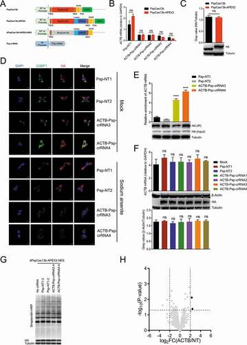
Figure 4. Identifying the RBPs of ACTB mRNA in cells inducibly expressing dPspCas13b-BioID2/TurboID/APEX2-NES
(A) Plasmids used in this figure. NES: nuclear export sequence; EGFP: enhanced green fluorescent protein; P2A: P2A self-cleaving peptide; HA: haemagglutinin tag.(B) HEK293T cells were co-transfected with PspCas13b or PspCas13b-APEX2/BASU/BioID2/TurboID and ACTB Psp-crRNA 4 to detect the mRNA level of ACTB after 48 hours.(C) Gray value analysis and western blot to measure the protein expression level of PspCas13b, PspCas13b-BioID2, PspCas13b-TurboID, PspCas13b-BASU and PspCas13b-APEX2.(D) Western blot to test the inducible expression ability and biotinylation activity of four cell lines inducibly expressing dPspCas13b- BioID2/TurboID/BASU/Apex2-NES. Different concentrations of doxycycline (Dox) were added to induce expression. Cells were collected together 27 hours after addition of doxycycline. For BioID2, biotin was added to the culture medium at a final concentration of 50 uM for 18 hours before harvesting cells; for TurboID, biotin was added at a final concentration of 500uM for 10 minutes before harvesting cells; for BASU, biotin was added at a final concentration of 200uM for 2 hours before harvesting cells; for APEX2, biotin-phenol was added at a final concentration of 500 uM for 30 minutes followed by a 1-minute exposure to 1 mM H2O2 before harvesting cells.(E) Timeline to capture the proteins that interact with ACTB mRNA using three cell lines inducibly expressing dPspCas13b-BioID2/TurboID/Apex2-NES. min: minute. h: hour.(F) Western blot to detect the expression level and biotinylation activity of cells collected from (E).(G) Volcano plots of ACTB mRNA-associated proteins identified by CBRPP in HEK293T cells inducibly expressing dPspCas13b-BioID2-NES. The significantly enriched proteins (P-value < 0.05 and fold change > 4) are shown as black dots (n = 3 for the ACTB Psp-crRNAs group and the non-targeting Psp-crRNAs group). Red dots represent known RBPs of ACTB mRNA in StarBase v2.0 database. Four previously uncharacterized RBPs of ACTB mRNA identified and validated in this study are shown as orange dots. FC: fold change, ACTB/NT: the ACTB Psp-crRNAs group/the non-targeting Psp-crRNAs group.(H) Left: RT-qPCR to measure the mRNA level of ACTB pulled down by anti-FLAG antibody in HEK293T cells transfected with indicated plasmids. EV: empty vector. Right: western blot to confirmed that proteins are specifically pulled down by anti-FLAG antibody not IgG. Results showed that some proteins found by CBRPP could significantly enrich ACTB mRNA compared with empty vector (EV) control.For B, C and H: n = 3, mean ± SD, two-sided Student’s t-test, ****P < 0.0001; ***P < 0.001; **P < 0.01; *P < 0.05; ns: not significant.
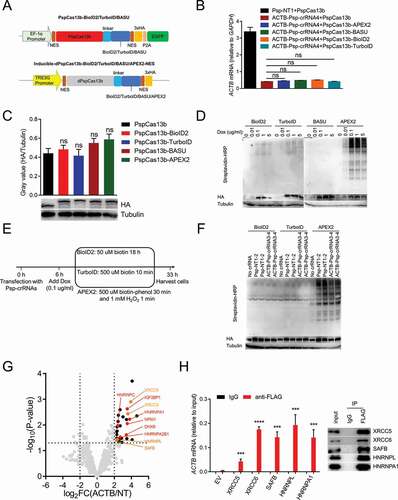
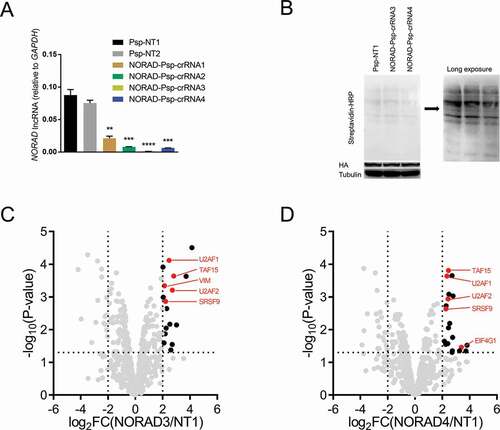
Figure 5. Identifying the RBPs of NORAD using CBRPP
(A) HEK293T cells were co-transfected with PspCas13b and NORAD Psp-crRNAs to detect the RNA level of NORAD after 48 hours.(B) Western blot to detect biotinylation activity in HEK293T cells inducibly expressing dPspCas13b-BioID2-NES transfected with NORAD Psp-crRNAs.(C-D) Volcano plots of NORAD-associated proteins identified by CBRPP in HEK293T cells inducibly expressing dPspCas13b-BioID2-NES. The significantly enriched proteins (P-value < 0.05 and fold change > 4) are shown as black dots (n = 3 for the NORAD Psp-crRNA 3 group, the NORAD Psp-crRNA 3 group and the non-targeting Psp-crRNA 1 group). Red dots represent known RBPs of NORAD in StarBase v2.0 database. FC: fold change. NORAD3/NT1: the NORAD Psp-crRNA 3 group/the non-targeting Psp-crRNA1 group. NORAD4/NT1: the NORAD Psp-crRNA 4 group/the non-targeting Psp-crRNA 1 group.For A: n = 3, mean ± SD, two-sided Student’s t-test, ****P < 0.0001; ***P < 0.001; **P < 0.01; *P < 0.05; ns: not significant.
Supplemental material