
Figures & data
Figure 1. Biochemistry of dihydrouridine.
A Reduction of uridine into dihydrouridine. B Dihydrouracil is a nonplanar nucleobase (carbon in gray, nitrogen in blue, oxygen in red and hydrogen in white). C First published structure of a ribonucleic acid (yeast tRNAAla) where D (red arrows) are shown in a loop at the 5’-end [Citation12]. D Schematic representation of ribose pucker. C5’ (orange dot) is considered as being above the C4’-O’-C1’ plane (red dashed line and red dots). Left panel: C3’-endo has the C3’ (green dot) above the plane. Right panel: C2’-endo has the C2’ (blue dot) above the plane. E C2’-endo pucker produces a longer 5’-phosphate/3’-phosphate distance and therefore spans the polynucleotide [Citation147]. F Schematic representation of ribose gauche-gauche and gauche-trans conformations. C5’ (purple dot) adopts different torsion angles (γ) that modulate the positioning of C5’-bound atoms (H5ʹ1,H5ʹ2 and O5’).
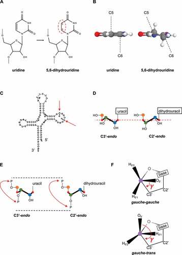
Table 1. Seminal studies on optical and structural properties of dihydro-uracil/-uridine
Figure 2. Molecular fate of D upon sodium borohydride (NaBH4) or alkaline (OH−) treatments.
R stands for ribose attached to the RNA chain. Red circles highlight ureido-groups.H+ stands for acid conditions. Details in the text.
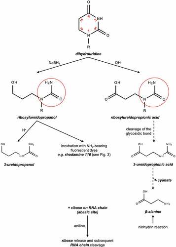
Table 2. Chemical reactions and techniques specifically applicable to D
Figure 3. Proposed mechanism of D rhodamine labeling following sodium borohydride (NaBH4) reduction.
Left panel, dihydrouridine is reduced to tetrahydrouridine that is characterized by an electrophilic carbonilamine (green circle), including the C4 hydroxyl group (C-OH). Upon addition of the nucleophilic rhodamine 110 in acid conditions, the tetrahydrocytidine intermediate is formed with its reactive Schiff base (blue circle). Covalent binding of rhodamine 110 occurs by substitution of the C4 hydroxyl group. Right panel, ureidopropanol generated by D-ring opening is replaced by rhodamine 110. R stands for ribose attached to the RNA chain.
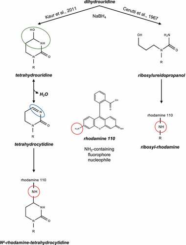
Figure 4. The biology of dihydrouridine.
A Specificity of yeast Dus enzymes for cytoplasmic tRNAs.B Specificity of E. coli Dus enzymes for tRNAs.C Putative enzymatic mechanism of Dus enzymes. Details in the text.D Currently available structures of Dus enzymes in different species (Sp.) from different domains (dm; B for Bacteria and E for Eukarya). The references are indicated in the right column.
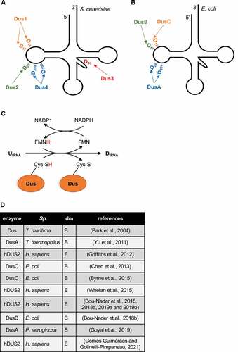
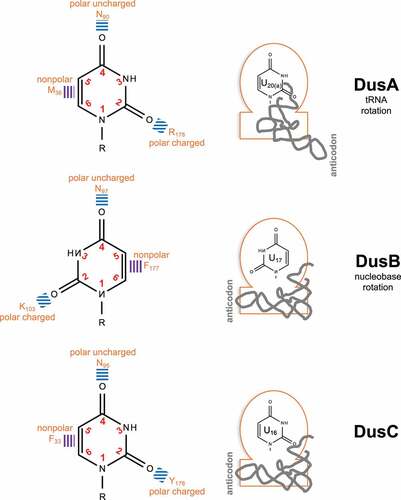
Figure 5. Molecular strategies of bacterial Dus enzymes for substrate specificities.
Left panel, interaction of the U nucleobase (in the context of the RNA chain (R)) with Dus amino acids (orange: F for Phe, N for Asn, K for Lys and Y for Tyr) through ionic (blue lines) or hydrophobic (purple lines) interactions. In the DusB catalytic pocket, the rotation of the U nucleobase (180°) is observed in comparison to the DusA and C counterparts. Right panel, schematic representations of the Dus enzymes (orange line) with the N-terminal nucleobase-containing catalytic domain (round) and the C-terminal RNA binding domain (rectangle). The tRNA (gray line) docking is similar in DusB and C but differs by a rotation (160°) in DusA. These strategies (tRNA or nucleobase rotations) allow the unvariable targeting of a specific uridine.
Supplemental material