
Figures & data
Figure 1. Two-step degradation reaction of pseudouridine. PUKI and PUMY are responsible for pseudouridine catabolism. Atomic numbering of the nucleobase is indicated.
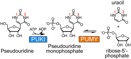
Table 1. Data collection and refinement statistics.
Figure 2. Structural features of the AtPUMY holoenzyme.
A. Monomeric structure of AtPUMY. The concave side of AtPUMY is shown, with the central β-sheet in blue. The figure orientations of A, B, and D are almost identical and corresponded to the left view of subunit A in (i.e., the front view of a trimer). Six α-helices (α1–α4, α10, and α13) were on the convex side, and α-helices α5–α9 and two β-strands were on the concave side. The putative Mn2+ ion and one sulphate ion are indicated by a black sphere and a space-filling model, respectively. The loop connecting β7, α7, and α8 is shown in magenta.
B. Zoom view of the metal-binding site in a monomer with an orientation in (A). Asp164 and a water molecule (green circle) across Asp164 are the axial ligands, and four water molecules are equatorial ligands. The dashed lines indicate possible coordination bonds to the metal ion and show a square bipyramidal geometry. Residues possibly stabilizing the binding of sulphate ion are indicated.
C. Front view of an AtPUMY trimer. Each subunit in the surface representation is coloured differently, and colour codes are maintained unless otherwise specified. The three-fold symmetry axis runs through the centre.
D. Active site of AtPUMY in a trimer. An α9* from the adjacent subunit (i.e., subunit C) participates in the active site of subunit A, which generates a pocket-shaped active site in the trimer. The black sphere represents the putative Mn2+ ion. The dashed line represents the Cα-interatomic distance between Arg116 at α5 and Asn286 at α12, which indicates the movement of α12 in response to the binding of ligands (see ).
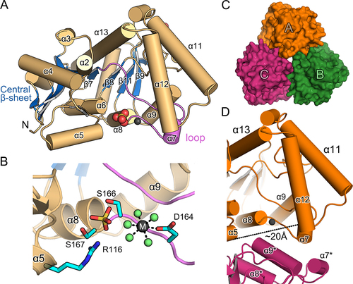
Figure 3. Ligand-binding modes and conformational changes in AtPUMY.
A. Citrate-binding mode in the AtPUMY(K185A)–Mn2+–citrate complex. This orientation is almost identical to that in . Citrate directly interacts with the Mn2+ ion, and residues in the vicinity of citrate, including the side chain of Thr289, are indicated. For clarity, possible hydrogen bonds and coordination bonds at the metal-binding site are indicated by solid lines, and α12 is absent from this presentation. An identical presentation in the presence of α12 is shown in Supplementary Fig. S4A.
B. ΨMP in the active site of the AtPUMY(K185A)–Mn2+–ΨMP/R5P complex. Residues, including the side chain of Thr289, within ~4 Å of ΨMP are indicated. Details are similar to those of (A). Note the water molecule between Thr149 and uracil-Ψ. An identical presentation in the presence of α12 is shown in Supplementary Fig. S4B.
C. R5P in the active site of AtPUMY(K185A)–Mn2+–ΨMP/R5P complex. R5P has a binding mode different from that of ΨMP. An equatorial water molecule was not identified in this structure. The presentation in the presence of α12 is also shown in Supplementary Fig. S4C.
D. Conformational changes in the active site. The lid elements of α11 and α12 underwent open-to-closed conformational changes in response to the binding of ligands, including citrate, R5P, and ΨMP. An open conformation (i.e., structure in grey) corresponds to the structure of the AtPUMY holoenzyme in ; the three subunits in the AtPUMY(K185A) – Mn2+–ΨMP complex represent the closed conformation (purple).
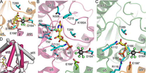
Figure 4. Enzymatic properties of AtPUMY.
A. Steady-state kinetic analysis of wild-type AtPUMY. Using ΨMP enzymatically synthesized as the substrate, the reaction product R5P was quantified using an enzyme-coupled assay. Each measurement was conducted in triplicate. Experimental details are provided in the Materials and methods.
B. AtPUMY activity towards C- and N-nucleosides. Reactions were performed as in (A) with 25 µM nucleoside 5’-monophosphates and a 60 s reaction. n.d., not detected.
C. Specific activities of mutant AtPUMYs towards 25 µM ΨMP. Reaction details are identical to (B), but enzyme concentrations were increased from 80 nM to 400 nM and 8 µM if no activity was detected. Each measurement was conducted in triplicate. n.d., not detected with 8 µM AtPUMY. Mutants were grouped according to their relative locations to each moiety in ΨMP (see ), and the inactive mutants were also grouped independently.
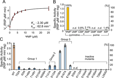
Table 2. Kinetic parameters of AtPUMY mutants.
Supplemental material
Supplemental Material
Download MS Word (1.5 MB)RHEE_8K06.pdf
Download PDF (3.2 MB)RHEE_8K05.pdf
Download PDF (1.2 MB)RHEE_8K07.pdf
Download PDF (2.7 MB)Data availability statement
The atomic coordinates and structural factors have been deposited in the Protein Data Bank (http://www.rcsb.org) under ID code 8K05, 8K06, and 8K07.