Figures & data
FIG. 1 The structure of balcalein (aglucone of balcalin). Adapted from Lai et al. (Citation2003).
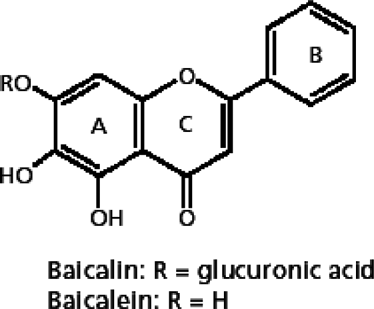
TABLE 1 The percentage of baicalein in isolated fractions of O. indicum
TABLE 2 Effect of the n- butanol fraction (100 mg/kg/day per os) of root bark of O. indicum on antibody (Ab) formation against SRBC in sensitized rats (humoral immune response)
TABLE 3 Effect of n- butanol fraction (100 mg/kg/day per os) of root bark of O. indicum on SRBC-induced hind paw edema in pre-sensitized rats
TABLE 4 Effect of the n- butanol fraction (100 mg/kg/day per os) of root bark of O. indicum on MDA content, anti-oxidant enzyme activities, and reduced glutathione levels
FIG. 2A Representative photomicrograph of splenic tissues recovered from rats on Day 22 of the respective indicated treatment regimens (WP = White pulp and RP = Red pulp)—Control rats. Left image at 10×, Right image at 40×.
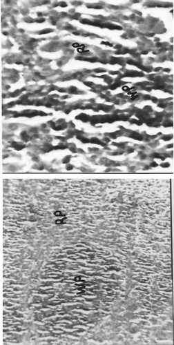
FIG. 2B Representative photomicrograph of splenic tissues recovered from rats on Day 22 of the respective indicated treatment regimens (WP = White pulp and RP = Red pulp)—n-Butanol extract-treated rats. Left image at 10×, Right image at 40×.
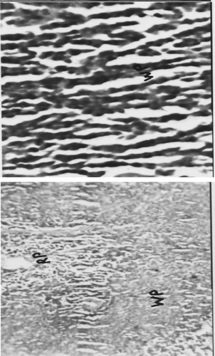
FIG. 2C Representative photomicrograph of splenic tissues recovered from rats on Day 22 of the respective indicated treatment regimens (WP = White pulp and RP = Red pulp)—Dexamethasone-treated rats. Left image at 10×, Right image at 40×.
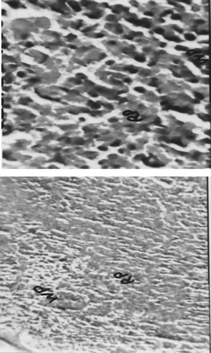
FIG. 3A Representative photomicrograph of thymus recovered from rats on Day 22 of the respective indicated treatment regimens (L = Lymphocytes, S = Sinusoid, HB = Hassel's Body. Left image at 10×, Right image at 40×.
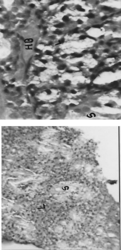
FIG. 3B Representative photomicrograph of thymus recovered from rats on Day 22 of the respective indicated treatment regimens (L = Lymphocytes, S = Sinusoid, HB = Hassel's Body. (B) n-Butanol extract-treated rats; Left image at 10×, Right image at 40×.
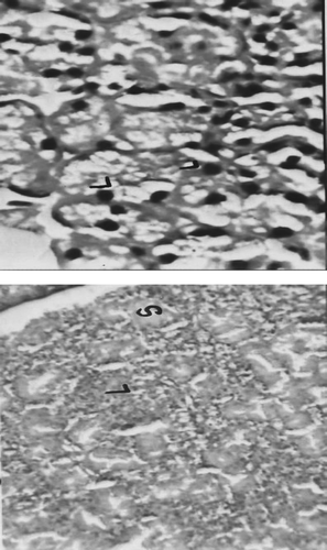
FIG. 3C Representative photomicrograph of thymus recovered from rats on Day 22 of the respective indicated treatment regimens (L = Lymphocytes, S = Sinusoid, HB = Hassel's Body. (C) Dexamethasone-treated rats. Left image at 10×, Right image at 40×.
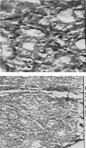
FIG. 4A Representative photomicrograph of axillary lymph node tissues recovered from rats on Day 22 of the respective indicated treatment regimens (C = Cortex, CP = Capsule, M = Medulla, and LF = Lymphoid follicles)—Control rats. Left image at 10×, Right image at 40×.
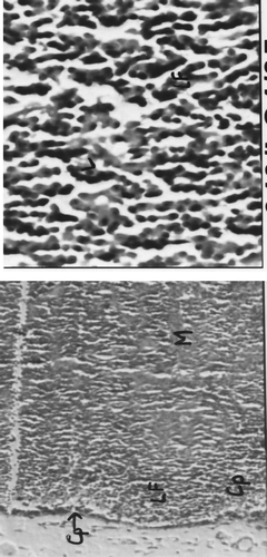
FIG. 4B Representative photomicrograph of axillary lymph node tissues recovered from rats on Day 22 of the respective indicated treatment regimens (C = Cortex, CP = Capsule, M = Medulla, and LF = Lymphoid follicles)—n-Butanol extract-treated rats. Left image at 10×, Right image at 40×.
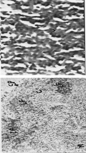
FIG. 4C Representative photomicrograph of axillary lymph node tissues recovered from rats on Day 22 of the respective indicated treatment regimens (C = Cortex, CP = Capsule, M = Medulla, and LF = Lymphoid follicles)—Dexamethasone-treated rats. Left image at 10×, Right image at 40×.
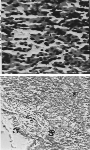
TABLE 5 Effect of the n-butanol fraction (100 mg/kg/day per os) of root bark of O. indicum on alum adjuvant-induced hind paw edema in pre-sensitized rats