Figures & data
FIG. 1 Experimental design to determine effects of developmental and early life exposure to TCE.
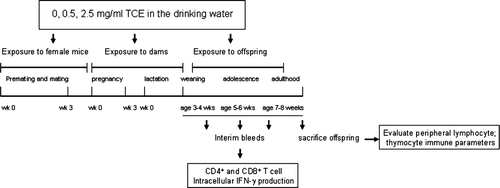
FIG. 2 TCE increased IFNγ produced by CD4+ and CD8+ T lymphocytes. (A) Individual offspring were bled at regular intervals following weaning to test for intracellular IFNγ production by peripheral blood CD4+ and CD8+ T-lymphocytes. After stimulation of whole blood with PMA and ionomycin for 5 hours, the cells were then 2-color stained with PE-anti-CD4 or PE anti-CD8 antibody in conjunction with FITC anti-IFNγ. The data represent the mean ± SD of CD4+ or CD8+ T-lymphocytes that were also positive for IFNγ. Results statistically different from control (* p < 0.05) or statistically different from 0.5 mg/ml TCE (** p < 0.05). (B) Representative dot plots from individual mice from the data presented in (5–6-wk timepoint) of IFNγ staining on CD4+ cells from mice exposed to water or TCE. (C) Equal numbers of spleen cells from the offspring in each treatment group were pooled for an n = 3 and the CD4+ T-lymphocytes were purified. The isolated CD4+ T-lymphocytes (1 × 10⁵ per well) were activated with plate-bound anti-CD3 antibody and soluble anti-CD28 antibody. Supernatants were collected after 72 hours incubation and analyzed for the presence of IL-2, IFNγ, IL-4, and TNFα using multiplex biomarker immunoassay kits with the Luminex 100 Bioanalyzer. The data were quantitated using standard curves and represented in the graphs as means ± SD in pg/ml.
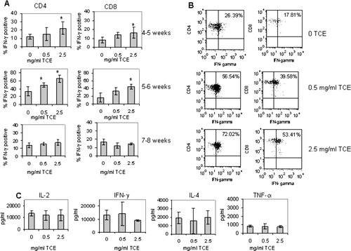
TABLE 1 Comparison of lymphocyte subpopulations in spleens of MRL+/+ mice exposed throughout development with TCE
FIG. 3 Increased IgG2a and anti-hostone levels in TCE-exposed offspring. Total IgG2a and IgG1 levels in the sera of individual mice were measured using standard ELISAs as described in Materials and Methods. Serum (diluted 1:100) was collected from the offspring at sacrifice and tested for IgG2a, IgG1, anti-histone, anti-ssDNA, or anti-dsDNA. The data are presented as the mean optical density (OD) + SD. Sera from aged adult MRL+/+ mice (8–10 months of age) were also tested as a control for positive responses for autoantibody production. *Statistically different from control (* p < 0.05).
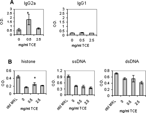
TABLE 2 Phenotypic alterations in the thymocyte profile of MRL+/+ mice exposed to TCE throughout development
FIG. 4 Percentage of thymocytes expressing CD4 and CD8. Thymocytes obtained from control and TCE-treated offspring were pooled for an n = 3 per treatment group. The cells were then subjected to staining with FITC anti-CD8 and PE anti-CD4 antibodies and examined by flow cytometry. The numbers within each dot plot represent the % of thymocytes that express CD4 and CD8.
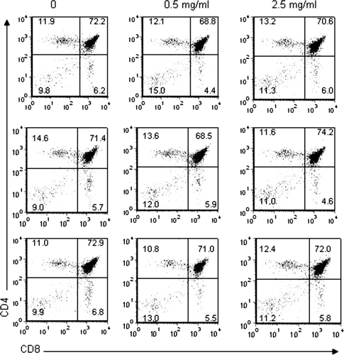
FIG. 5 TCE exposure inhibited apoptosis of DN thymocytes isolated from MRL+/+ offspring. Thymocytes obtained from control and TCE-treated offspring were pooled from each treatment group for a statistical n = 4 and were cultured in medium for 24 hours (2 × 10⁶/ml). The apoptotic cells were 3-color stained with FITC Annexin V, PE-anti-CD8 and biotinylated anti-CD4 followed by PerCP streptavidin and examined by flow cytometry. The graphs represent the mean ± SD Annexin V staining from gated SP, DN, or DP populations. *Significantly different from control (p < 0.05).
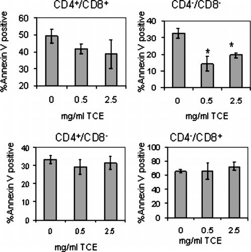
FIG. 6 TCE did not alter AICD of peripheral T-lymphocytes. Spleen cells isolated form pooled samples within each treatment group (n = 3) were cultured at a concentration of 2 × 10⁶ per ml and activated to undergo apoptosis as described in Materials and Methods. The cells were harvested and three-color stained with PE-anti-CD8, FITC Annexin V, and biotinylated anti-CD4 and PerCP streptavidin. Presented are representative FITC histograms after gating on the PE+ or PerCP+ cells. The percentage (%) within each histogram refer to the % Annexin V positive CD4+ or CD8+ T-lymphocytes treated with anti-CD3 (grey histogram). The black histograms in each plot represent Annexin V staining of CD4 or CD8 T-lymphocytes in the absence of anti-CD3 stimulation.
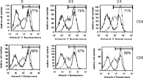
FIG. 7 TCE exposure did not affect litter size but promoted decreased growth in the offspring. (A) Three female mice per treatment group produced litters containing similar numbers of pups. The bars in the graph represent the mean ± SD number of pups delivered per treatment group. (B) pup was weighed once weekly from the time immediately following weaning until sacrifice. The symbols in the graph represent mean ± SD of the body weights from each group. Results are significantly different from control (* p < 0.05).
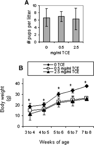
TABLE 3 Water consumption