Figures & data
TABLE 1 Protocols of sensitization, challenge and measurements for each group
FIG. 1 Penh values in pre-challenge, challenge, and post-challenge periods. Conscious mice were individually held in a restrained flow whole body plethysmograph and enhanced pause (Penh) was recorded every 1 min during 5 min pre-challenge, 15 min challenge, and 40 min post-challenge periods using an analytical recording system. Mice were challenged with fine TMA dust. Symbols represent airway responses of 6 mice/group.
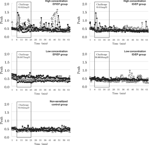
TABLE 2 Enhanced pause (Penh) before, during and after challenge
FIG. 2 Total serum IgE values in each group. Two days after inhalation challenge, 6 mice/group were anesthetized with pentobarbital and blood samples drawn from the posterior vena cava. Total serum IgE values were measured with a sandwich enzyme-linked immunosorbent assay (ELISA). ** p < 0.01 compared with non-sensitized control mice.
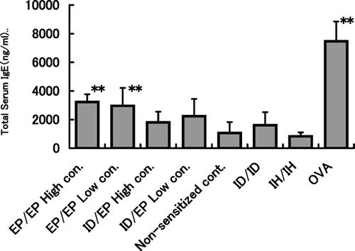
TABLE 3 BALF analyses in the EP/EP, ID/EP and Non-sensitized control groups
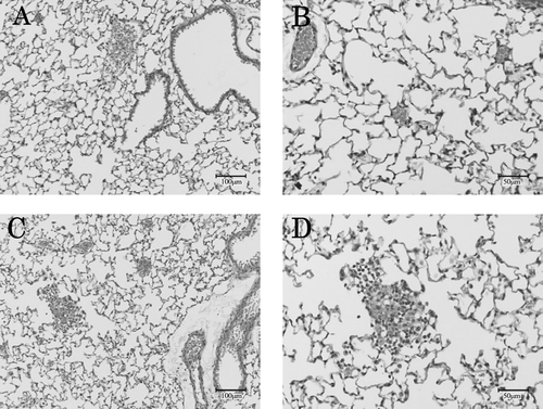
FIG. 3 Representative pulmonary region of lungs. Pulmonary regions from non-sensitized control group (A, B) and from EP/EPhi group (C, D) 48 hr after challenge with TMA dust. A cell infiltration (including eosinophils) into the airspace and perivascular portions was observed only in TMA-sensitized and -challenged mice.