Figures & data
FIG. 1 Synchronous fluorescence spectrophotometry for determining adduction of BPDE to DNA. Result of one representative experiment is shown. Maximum excitation wavelengths for tetraols are 349 to 352 nm, and the wavelength remains high at 355 to 375 nm.
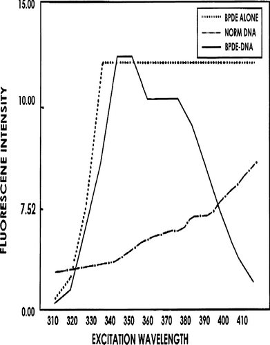
FIG. 2 Detection of BPDE-DNA adducts in MACS-isolated CD4+CD8+ DP and CD4+ CD8− SP cells from thymus of B(α) P- and corn oil (CO)-exposed progeny (1-wk-of-age). Data shown are representative of 4 experiments. X-axis: Intensity of Green Fluorescence. Y-axis: Intensity of Red Fluorescence. Results presented are from: (A) CO thy cells only; (B) B(α) P thy cells only; (C) CO cells + propidium iodide (PI) + rabbit anti-BPDE-DNA 1° Ab + FITC-goat anti-rabbit IgG 2° Ab; (D) B(α) P cells + PI + 1° Ab + 2° Ab; and (E) B(α) P thy cells + PI + non-specific rabbit IgG + 2° Ab (to reflect non-specific binding of 2° Ab). Numbers in Quadrant 2 are percentage of cells detected containing BPDE-DNA adducts. For example, in , 18% of cells stained with PI (DNA-red) and rabbit anti-BPDE-DNA FITC (green), indicating presence of BPDE-DNA in progeny cells.
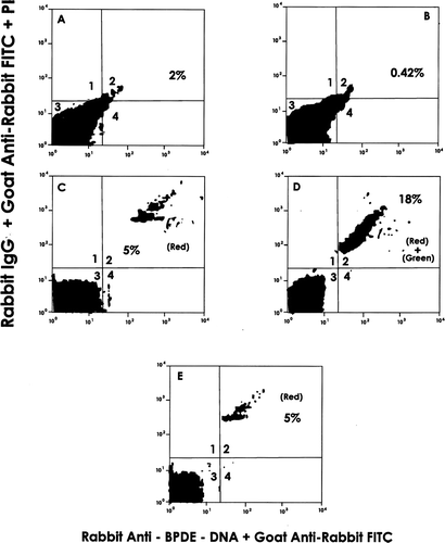
FIG. 3 Detection of BPDE-DNA adducts in splenic CD4+CD8− spleen cells. Mini-macs-isolated cells were treated with saponin + PI + rabbit anti-BPDE-DNA IgG + FITC-goat anti-rabbit IgG. Data shown are representative of 3 experiments.
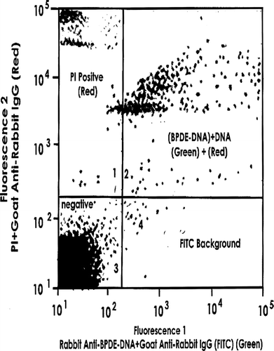
FIG. 4 Detection of BPDE-DNA in CD4+CD8− fetal liver cells via [32P]-post-labelling. Data are representative of 3 experiments. Arrow #1 points to BPDE-DNA complex and Arrow #2 to origin. Fetal liver samples of (A) Corn oil (CO) control and (B) B(α) P-exposed progeny collected at Day 17 gestation.
![FIG. 4 Detection of BPDE-DNA in CD4+CD8− fetal liver cells via [32P]-post-labelling. Data are representative of 3 experiments. Arrow #1 points to BPDE-DNA complex and Arrow #2 to origin. Fetal liver samples of (A) Corn oil (CO) control and (B) B(α) P-exposed progeny collected at Day 17 gestation.](/cms/asset/0fbc9c1a-6e0a-4a81-a731-1ba0d29227f2/iimt_a_267861_uf0004_b.gif)
FIG. 5 One-way allogeneic MLR by isolated SP CD4+ thymus cells that do or do not express the BPDE-DNA adduct. Values shown represent mean percent of corn oil mouse [control] MLR activity (± SD) from 4 experiments. Control thymus cell MLR CPM = 25 × 103. Difference between relative activities of BPDE-DNA+ and BPDE-DNA− cells was significant (p < 0.001).
![FIG. 5 One-way allogeneic MLR by isolated SP CD4+ thymus cells that do or do not express the BPDE-DNA adduct. Values shown represent mean percent of corn oil mouse [control] MLR activity (± SD) from 4 experiments. Control thymus cell MLR CPM = 25 × 103. Difference between relative activities of BPDE-DNA+ and BPDE-DNA− cells was significant (p < 0.001).](/cms/asset/3aa6d127-8c16-4e5f-ba4e-f2784ba1ff87/iimt_a_267861_uf0005_b.gif)