Figures & data
Figure 1. Survival of ICR and C3H/HeJ mice administered SPG/IND. (A) Eight-week-old ICR mice (saline/IND; n = 6, SPG/IND; n = 5) were administered SPG (100 mg/mouse) intraperitoneally (IP) on Days -5, -3, and -1, and indomethacin (IND; 5 mg/kg) per os from days 0 to 14. Mortality was monitored. (B) Eight-week-old C3H/HeJ mice (saline/IND; n = 14, SPG/IND; n = 15) were administered SPG (100 mg/mouse) or saline IP on Days -5, -3, and -1, and indomethacin (IND; 5 mg/kg) per os from Days 0 to 14. Mortality was monitored.
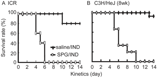
Figure 2. GOT and GPT levels in sera from SPG/IND-treated C3H/HeJ mice. Eight-week-old C3H/HeJ mice (saline/IND, n = 4; SPG/IND, n = 3) were administered SPG (100 μg/mouse) or saline IP on Day -5, -3, and -1, and indomethacin (IND, 5 mg/kg) per os from Day 0 to 7. Sera were prepared on Day 5 and 7. GOT and GPT levels were measured with kits using sera obtained on Day 7 (serum of one mouse in saline/IND group could not be obtained, thus used serum on Day 5). Results shown are the mean (± S.D) Karmen activity levels; a karmen unit is a formerly-used expression of aminotransferase activity (based on a 0.001 change in absorbance of NADH/min). Significance (**p < 0.05, ***p < 0.01), was evaluated using a student’s t-test.
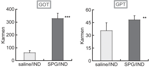
Figure 3. Effect of Pam3 or CpG on IL-6 production of splenocytes and peritoneal exudate cells. (A, B) Spleen cells were collected from naive C3H/HeJ mice, and (C, D) peritoneal exudates cells (PEC) were collected from thioglycolate-broth administered C3H/HeJ mice. Cells were incubated with Pam3 or CpG for 24 hr at 37°C in 5% CO2 (a, c; Pam3, b, d; CpG). IL-6 presence in the supernatant was measured by ELISA. Results are shown as the mean ± SD.
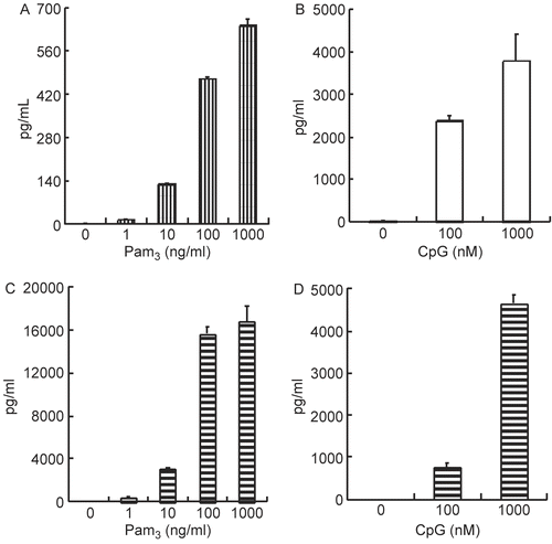
Figure 4. Synergistic effect of Pam3/CpG on IL-6 production of splenocytes and peritoneal exudates cells. (A) Spleen cells were collected from naive C3H/HeJ mice, and (B) peritoneal exudates cells (PEC) were collected from thioglycolate-broth administered C3H/HeJ mice. Cells were incubated with Pam3 in the presence or absence of CpG for 24 hr at 37°C in 5% CO2. IL-6 presence in the supernatant was measured by ELISA. Results are shown as the mean ± SD.
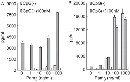
Figure 5. CYP3A11 protein expression in liver microsomes from SPG-administered mice. Five-week-old ICR mice (A) and eight-week-old C3H/HeJ mice (B) were administered SPG (100 μg/mouse) or saline IP on Days -5, -3, and -1. On Day 0, liver microsomes were obtained and CYP3A11 protein expression was measured by Western blotting. Lane 1, 500 μg/ml; Lane 2, 250 μg/ml; Lane 3, 125 μg/ml; Lane 4, 62.5 μg/ml; Lane 5, 31.25 μg/ml; Lane 6, 15.625 μg/ml.
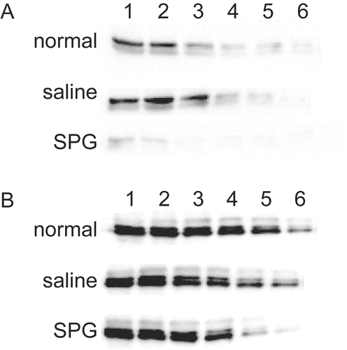
Figure 6. CYP3A11 protein expression in liver microsomes from SPG/IND-administered mice. Five-week-old ICR mice (A) and eight-week-old C3H/HeJ mice (B) were administered SPG (100 μg/mouse) or saline IP on Days -5, -3, and -1, and IND (5 mg/kg) per os from Day 0 to 2. On Day 2, liver microsomes were obtained and CYP3A11 protein expression was measured by Western blotting. Lane 1, 500 μg/ml; Lane 2, 250 μg/ml; Lane 3, 125 μg/ml; Lane 4, 62.5 μg/ml; Lane 5, 31.25 μg/ml; Lane 6, 15.625 μg/ml.
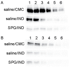
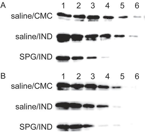
Figure 7. CYP3A11 protein expression of liver microsomes from SPG/IND-administered mice just before disease. Five-week-old ICR mice (A) and eight-week-old C3H/HeJ mice (B) were administered SPG (100 μg/mouse) or saline IP on Days -5, -3, and -1, and indomethacin (IND, 5 mg/kg) per os from Day 0 to 4. On Day 3 and 4, liver microsomes were obtained and CYP3A11 protein expression was measured by Western blotting. Lane 1, 500 μg/ml; Lane 2, 250 μg/ml; Lane 3, 125 μg/ml; Lane 4, 62.5 μg/ml; Lane 5, 31.25 μg/ml; Lane 6, 15.625 μg/ml.
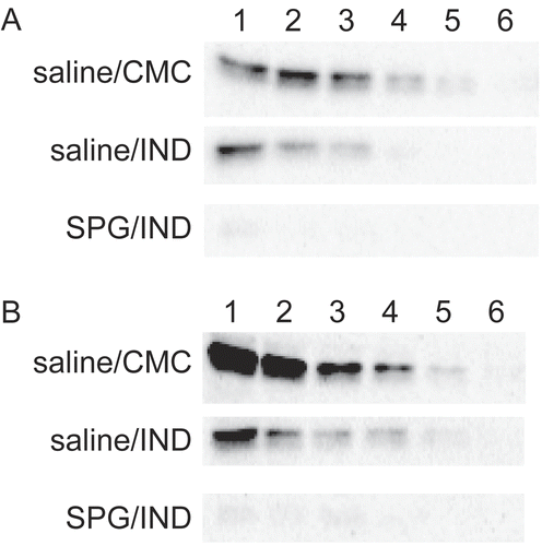
Figure 8. CYP2E1 protein expression in liver microsomes from SPG-administered mice. Five-week-old ICR mice (A) and eight-week-old C3H/HeJ mice (B) were administered SPG (100 μg/mouse) or saline IP on Days -5, -3, and -1. On Day 0, liver microsomes were obtained and CYP2E1 protein expression was measured by Western blotting. Lane 1, 500 μg/ml; Lane 2, 250 μg/ml; Lane 3, 125 μg/ml; Lane 4, 62.5 μg/ml; Lane 5, 31.25 μg/ml; Lane 6, 15.625 μg/ml.
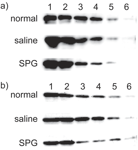
Figure 9. CYP2E1 protein expression in liver microsomes from SPG/IND-administered mice. Five-week-old ICR mice (A) and 8-week-old C3H/HeJ mice (B) were administered SPG (100 μg/mouse) or saline IP on Days -5, -3, and -1, and indomethacin (IND, 5 mg/kg) per os from Day 0 to 2. On Day 2, liver microsomes were obtained and CYP2E1 protein expression was measured by Western blotting. Lane 1, 500 μg/ml; Lane 2, 250 μg/ml; Lane 3, 125 μg/ml; Lane 4, 62.5 μg/ml; Lane 5, 31.25 μg/ml; Lane 6, 15.625 μg/ml.
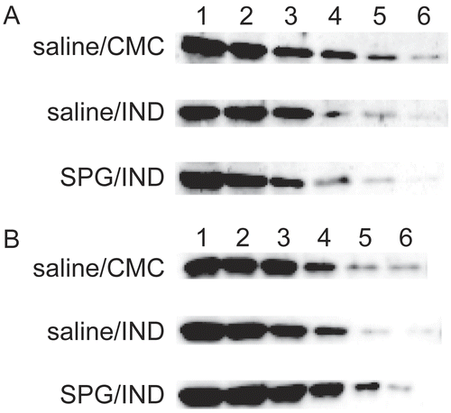
Figure 10. CYP2E1 protein expression in liver microsomes from SPG/IND-administered mice just before disease. Five-week-old ICR mice (A) and 8-week-old C3H/HeJ mice (B) were administered SPG (100 μg/mouse) or saline IP on Days -5, -3, and -1, and indomethacin (IND, 5 mg/kg) per os from Day 0 to 4. On Days 3 and 4, liver microsomes were obtained and CYP2E1 protein expression was measured by Western blotting. Lane 1, 500 μg/ml; Lane 2, 250 μg/ml; Lane 3, 125 μg/ml; Lane 4, 62.5 μg/ml; Lane 5, 31.25 μg/ml; Lane 6, 15.625 μg/ml.