Figures & data
Figure 1. Experimental protocol used to sensitize and challenge BALB/c mice with TMA in acetone/olive oil solvent. For the sensitizing protocol, three different schedules were tested as follows; 1-wk sensitization (A) (Days 1–3 dermal sensitization and Day 17 dermal challenge), 2-wk sensitization (B) (Days 1–3, 8–10 dermal sensitization and Day 24 dermal challenge) and 3-wk sensitization (C) (Days 1–3, 8–10, 15–17 dermal sensitization and Day 31 dermal challenge). A 25 μl aliquot of TMA solution or solvent only was applied to the dorsum of each ear of each mouse for dermal sensitization. Mice were then challenged 2 wk after last sensitization. The same volume of TMA solution or solvent only was applied to the dorsum of each ear of each mouse for dermal challenge.
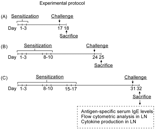
Table 1. Experimental groups used in this study.
Figure 2. TMA-specific IgE levels in mouse serum isolated 1 d after challenge with TMA solution or solvent alone. Experimental groups are as in . Results (circles) are values for (titer); bar indicates mean titer value. Statistical significance is marked by asterisks: *p < 0.05, **p < 0.01 (Tukey’s t-test).
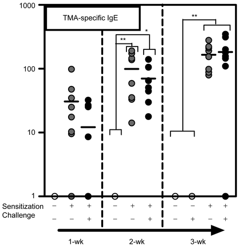
Figure 3. Total numbers of lymphocytes (A), numbers of B220+ IgE+ cells (B), and numbers of B220+ MHC Class II+ cells (C) in auricular lymph nodes from mice treated with TMA solution or solvent alone. Dot-plots of B220+ IgE+ cells in the +/+ treatment group receiving a 1-wk, 2-wk or 3-wk sensitization (D) are also shown. Circles show the staining pattern of B220+ IgE+ cells. Experimental groups are as in . Results are expressed as number of cells (mean ± SD). Statistical significance is marked by asterisks: *p < 0.05, **p < 0.01 (Tukey’s t-test).
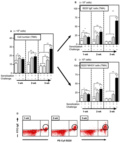
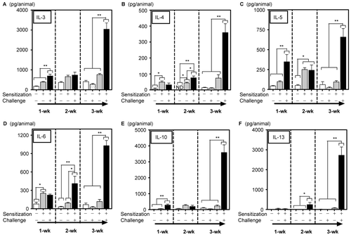
Figure 4. Cytokine production (A: IL-3, B; IL-4; C: IL-5; D; IL-6, E: IL-10, F: IL-13) by auricular lymph node calls of mice treated with TMA solution or solvent. To determine the effects on cytokine production, lymph node cell suspensions were cultured with T-cell antibodies (CD3 and/or CD28) for 24 or 96 hr (see details of culturing conditions for specific cytokines in Methods), and the supernatant was assayed by cytometric bead array. Experimental groups are as in . Results are expressed as mean (pg/animal) levels ± SD. Statistical significance is marked with asterisks: *p < 0.05, **p < 0.01 (Tukey’s t-test).