Figures & data
Table 1. Baseline characters table for 451 preeclamptic placentas (PIH) with different types of vasculopathy and associated pathologic findings. The baseline character table with p value calculation is generated by using the R-statistics program as described. C: C-section delivery; V: Vaginal delivery; cHTN: chronic hypertension; MFI: maternal floor infarction; Cord issues include marginal or velamentous insertions, 2 vessel cord, true knots, Others include oligohydramnios, polyhydramnios, placental previa, placental increta/percreta, bilobed placentas, fetal anomalies, chromosomal abnormalities, maternal history of IBDs, lupus, thyroid diseases, cholestasis and cancers.
Table 2. Baseline characters table for 583 placentas from non-preeclampsia patients with different types of vasculopathy and associated pathologic findings. The baseline character table with p value calculation is generated by using the R-statistics program as described.
Table 3. Baseline characters table for 877 placentas with vasculopathy associated with or without preeclampsia. The baseline character table with p value calculation is generated by using the R-statistics program as described.
Table 4. Placental weight associated with vasculopathy and other pathologic features to assess the effect of individual pathologic features on placental weight development. All placentas with vasculopathy are included (n = 877), and placentas associated with preeclampsia but not vasculopathy are excluded (n = 157).
Figure 1. Morphologic features of maternal spiral artery remodeling in early implantation site with immunohistochemical staining for CD56, pancytokeratin (AE1/AE3) and CD68 expression. (A) Implantation site of early missed abortion with hematoxylin & eosin (H&E) stain. (B–D) The same section of implantation site as panel A with CD56, AE1/AE3 and CD68 immunostaining. (A–D at 400× magnification). Arrow indicates weak CD68 reactivity.
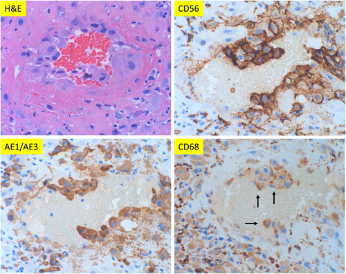
Figure 2. Morphologic features of classic decidual vasculopathy of preeclamptic placentas from the decidua basalis with immunohistochemical staining for CD56, AE1/AE3 and CD68 expressions. (A) Classic decidual vasculopathy with acute atherosis and fibrinoid medial necrosis by hematoxylin & eosin (H&E) stain. The same section of placenta as in H&E panel with CD56, AE1/AE3 and CD68 immunostaining. (All at 400× magnification). Arrows indicate positive reactivity to individual markers. (B) Decidual vasculopathy with immunostaining for CD56 and GATA3. Partial involvement of spiral artery wall by fibrinoid medial necrosis and acute atherosis in panel H&E stain. Adjacent space is an endometrial gland. The same vasculopathy as in H&E panel with immunostaining for CD56 and GATA3 (nuclear signal). All at 200× magnification.
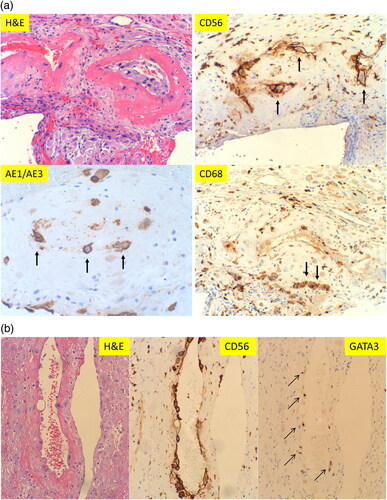
Figure 3. Vasculopathy in the decidual capsularis (fetal membrane roll) with immunostaining for CD56, CD68 and (pancytokeratin) AE1/AE3. Two separate vessels are shown in H& E with corresponding immunostaining. All sections are at 200× magnification.
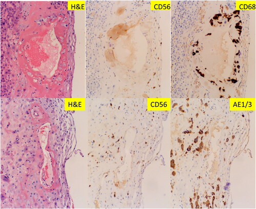
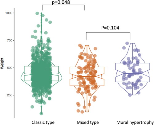
Figure 4. Placental weight comparisons between the four types of vasculopathy: Classic type (including acute atherosis and fibrinoid medial necrosis) (n = 198), mural hypertrophy (n = 59), mixed type (mixed classic type and mural hypertrophy) (n = 37) and no vasculopathy (n = 157) in placentas from preeclampsia (PIH). P-value was calculated by using the ANOVA test in R-statistics program.
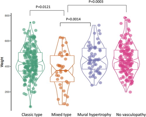
Figure 5. Correlation in aggregates of placental weight, gestational age and various types of decidual vasculopathy in patients with preeclampsia including early onset and late onset preeclampsia using conditioning plot (R-statistics package). Classic type n = 198, mural hypertrophy n = 59, mixed type n = 37, no vasculopathy n = 157. Details of specific type of vasculopathy and placental weight correlation is shown in the subsequent figures and tables.
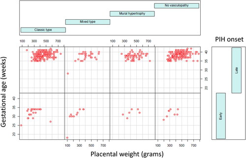
Figure 6. Placental weight comparison from patients with early onset preeclampsia (n = 60) versus late onset preeclampsia (n = 391) (left panel) and C-section delivery (n = 227) versus vaginal delivery (n = 224) (right panel). Early onset preeclampsia (n = 60) represents 13.3% of total preeclampsia in current study. The baseline characters are listed in .
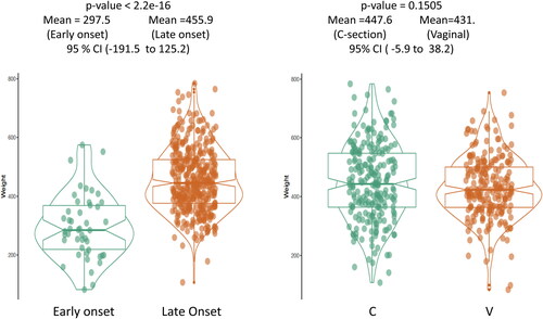
Figure 7. Placental weight comparison from patients with vasculopathy but no preeclampsia (PIH) or hypertensive disorders. The baseline characters are listed in . Classic type vasculopathy n = 504, Mixed type vasculopathy n = 75, mural hypertrophy n = 4.
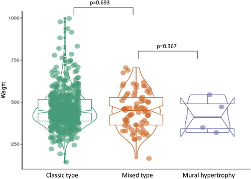
Figure 8. Placental weight comparison for all placentas with vasculopathy associated with or without preeclampsia. Classic type vasculopathy n = 702, mixed type vasculopathy n = 112, mural hypertrophy n = 63. The baseline characters are listed in .