Figures & data
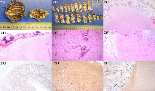
Figure 1. (A) The infant’s left forehead and upper eyelid were wine colored, which was light red and smooth. (B) The sulci and fissure of the bilateral cerebral hemispheres were widened and deepened, and the gyri were narrowed. The left cerebral hemisphere was more evidently atrophied than the right, and bilateral lateral ventricles and third ventricles were widened. (C) Diffuse gyri-like enhancement was observed on the brain surface of the left cerebral hemisphere, and linear enhancement was observed on the brain surface of the right parietal lobe.
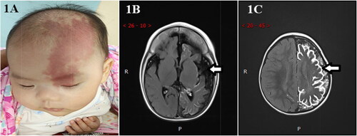
Table 1. Clinicopathological data of 10 children with Sturge–Weber syndrome.
Figure 2. (A) Gross examination of the meningeal surface reveals a network of small grayish-red vessels. (B) The boundary between the cortex and white matter was not clear on the parenchymal section of the brain, and the cortex was gray-red and tough, with many brown-yellow hard calcifications. (C) The leptomeningeal angiomatosis were thickened on the surface of brain tissue, and irregular dilated capillary lumens were widely distributed in the subarachnoid space, presenting a cavernous hemangiomatous change, H&E 10 × 4. (D) Malformed vessel wall showed obvious calcification, HE 10 × 4. (E) Neuronal cells proliferation are seen around calcification in the superficial brain parenchyma, H&E 10 × 10. (F) Cerebral cortical atrophy H&E 10 × 4. (G) NeuN showed disordered cortical structure around leptomeningeal angiomatosis and partial neuron loss, IHC 10 × 4. (H) NF showed that the cytoplasm of individual neurons was highly stained, IHC 10 × 4. (I) Endothelial cell CD31 positive expression of IHC 10 × 4