
Figures & data
Figure 1. Brain Bard Monopty needle with brain core.

Figure 2. Procedure for collection of brain cores from the anterior and posterior fontanel.
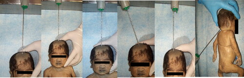
Table 1. Gestational age and head circumference of newborns.
Table 2. Histopathological findings of MITS from anterior and posterior fontanel.
Figure 3. Histopathological findings (a) Subarachnoid hemorrhage, (b) Parenchymal microhemorrhage with anoxic neurons, (c) Periventricular area with germinal matrix with congestion, (d) Meningitis, (e) Cerebellum and (f) Choroid plexus hemorrhage.
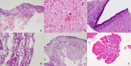
Table 3. Findings of MITS and CDA.
Table 4. Sensitivity, specificity, PPV, NPV, and associated 95% confidence interval of MITS when compared to CDA.
Data availability statement
The datasets used and analyzed during the current study are attached in the documents. Furthermore, information will be available from the corresponding author upon reasonable request.