Figures & data
Figure 1. A schematic overview of cranial development from the neural crest. Left: The dark yellow structure (NT) illustrates the neural tube. The notochord (n) is drawn as a red line passing through the vertebral corpora. The green structures are the facial bones developed from the neural crest. Four segments of neural crest cells are schematically demonstrated. These cells arise from the rim of the neural crest, marked by green dots (a, b, c, d), and from here, they follow the pathways (arrows) toward the anterior aspects of the face. The yellow structures illustrate the hemispheres, and below the hemispheres is the cerebellum. Center: This figure is a schematic drawing of the left illustration seen from behind. The notochord is a red line (n) and the dark yellow (NT) is the neural tube. Note that the four green dots (a, b, c, and d) appear on the right side and also on the left side of the neural tube. This is important for the understanding of unilateral malformations only involving the right or left neural crest. The arrows from the green dots indicate a pathway for the migrating cells to the jaws. Right: A seemingly normally developed perinatal cranium, seen from top to bottom. The cranium might be slightly deformed during storage and appear slightly asymmetrical. As this is a perinatal cranium, the teeth have not erupted. In the posterior view, Wormian bones appear, and the occipital squama appears divided into a lower and an upper bulging part. The upper part has a desmal origin, developed like the parietal bones. The lower part has a cartilaginous origin, comparable to the cartilaginous development of arches in the vertebrae.
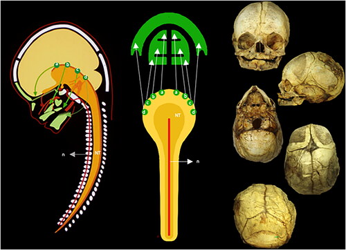
Figure 2. Illustration of the neural crest cells schematically transferred from experimental animals to a radiograph from a child 10 years of age. Left: This figure can be compared to the left drawing in . The colors demonstrate different fields. The yellow is the frontonasal field (A). The red is the maxillary field (B). The orange is the palatine field (C). The blue color (D) is the mandibular field. Not formed by neural crest cells are the theca field in purple (E) and the occipital field in green (O). The occipital field arises, like the vertebral bodies, from cartilage, influenced by the notochord. Right: This figure indicates the anterior view of the cranium. The colors and letters are comparable to the colors and letters in the left figure. Inserted figure between the left and right: This drawing indicates the well-known developmental fields marked by body lines. For a long time, the developmental fields in the head were not known (absence of lines) The neural crest cell migration in different directions explained the cranial development (Le Douarin [Citation12, Citation13]).
![Figure 2. Illustration of the neural crest cells schematically transferred from experimental animals to a radiograph from a child 10 years of age. Left: This figure can be compared to the left drawing in Figure 1. The colors demonstrate different fields. The yellow is the frontonasal field (A). The red is the maxillary field (B). The orange is the palatine field (C). The blue color (D) is the mandibular field. Not formed by neural crest cells are the theca field in purple (E) and the occipital field in green (O). The occipital field arises, like the vertebral bodies, from cartilage, influenced by the notochord. Right: This figure indicates the anterior view of the cranium. The colors and letters are comparable to the colors and letters in the left figure. Inserted figure between the left and right: This drawing indicates the well-known developmental fields marked by body lines. For a long time, the developmental fields in the head were not known (absence of lines) The neural crest cell migration in different directions explained the cranial development (Le Douarin [Citation12, Citation13]).](/cms/asset/15bc734a-5bbe-43cd-8957-4392d31d77dd/ipdp_a_2338434_f0002_c.jpg)
Figure 3. An overview of the five perinatal crania is analyzed from row 1 to row 5. Two crania have mandibular bones, and three do not have mandibular bones. Each cranium is demonstrated from top to bottom in the cranial base view, the thecal view, the anterior view, the lateral view, and the posterior view. Cranium 1, Row 1: A large malformation (absence of occipital bone), marked green in , is observed in the occipital view. In the thecal view, the absence (or early closure) of fontanel is apparent. In the frontal view, a low appearance of the diminutive frontal bones appears. In the posterior view, an absence of bone appears below the lambdoid suture. Cranium 2, Row 2: Absence of bone in the middle of the face (yellow area in ) and presence of septum nasi (arrow) appear in the occipital view. In the thecal view, a diminutive frontal fontanel appears. In the frontal view, the mid-axial cleft does not affect the orbital structures. In the lateral view, a short arcus zygomaticus can be observed. In the posterior view, an extra triangular bone appears above the occipital squama. Cranium 3, Row 3: In this cranium, only the mid-axial part of the face appears (this is the yellow part that was missing in Cranium 2). The regions present are the red and orange fields in . The nasal septum is marked by an arrow. In the thecal view, the fontanel appears normal. In the frontal view, the cavum nasi appears diminutive, and the lower rim of the orbita is absent. In the lateral view, the zygomatic bone and the arcus zygomaticus are missing. Cranium 4, Row 4: A narrow and long cranium appears from the occipital view. The absence of zygomatic bone and arcus is observed (orange areas in ). In the thecal view, the posterior part of the parietal bones is fused (synostosis). In the anterior part, the interfrontal bones are separated by a broad suture. Irregularities in the suture system also appear in the posterior view. Cranium 5, Row 5: In the occipital view, the palate appears narrow posteriorly. From the thecal view, parts of the frontal bone are visible anteriorly, and parts of the occipital bone are visible in the posterior direction. There is no thecal bone (purple area in ) uniting these two parts of the skull. In the frontal view, a narrow interocular distance is observed. In the lateral view, the maxillary complex appears diminutive and retrognathic. In the posterior view, there appear to be fusions between the cervical vertebrae. Bilateral malformations in the lower part of the occipital squama are observed.
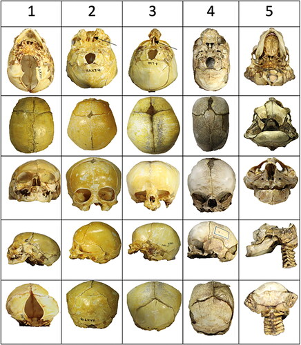
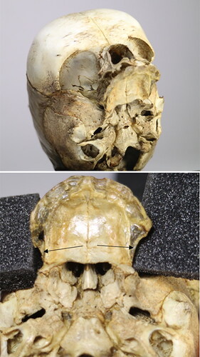
Figure 4. A comparison of cranium 2 () diagnosed holoprosencephaly, mid-line cleft type without facial midaxial structures and cranium 3 eventually diagnosed with Threscher Collin syndrome with midaxial facial structures, but absence of the lateral facial structures. Both specimens have a septum nasi (arrows) which proves that the septum does not develop from the neural crest but from the anterior cartilaginous cranial base. A comparison of the two specimens demonstrates that the orbital cavities are normal in case 2 but abnormal in case 3, lacking the zygomatic bone.
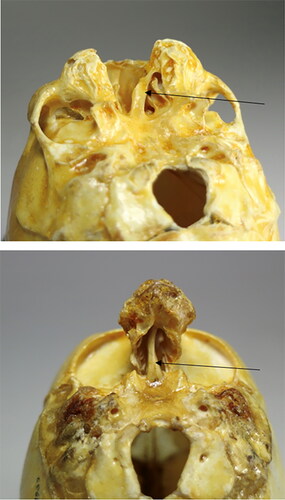
Figure 5. Two specific views on specimen 4 () were eventually diagnosed as scaphocephalic. The upper figure demonstrates an absence or malformation in the zygoma region and the vertical part of the palatal bone. The lower part illustrates, in a palatal view, the absence of alveolar bone in the maxillary molar region (arrows). The teeth are not visible. The timing between the tooth formation and the alveolar bone formation could be interrupted, but this cannot be concluded.