Figures & data
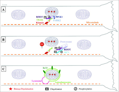
Figure 1. Model for local mitophagy of damaged mitochondria in neuronal axons. (A) Damage to axonal mitochondria leads to the accumulation of PINK1 and PARK2 on the mitochondrial outer surface where they interact with the motor complex composed of the RHOT/Miro and TRAK/Milton adaptor proteins and the kinesin motor protein. (B) PINK1- and PARK2-mediated proteasomal degradation of RHOT arrests both anterograde and retrograde motility of these organelles. This, and the parallel degradation of MFN1/2, are an early action of the PINK1 and PARK2 pathway prior to mitophagy and may help quarantine damaged mitochondria by preventing their fusion with healthy neighbors. (C) Accumulation of PARK2 on the stationary damaged mitochondria promotes their local engulfment within LC3-positive autophagosomes. Mitophagosomes fuse with axonal lysosomes to form autolysosomes and their mitochondrial content is degraded locally within the axon.