Figures & data
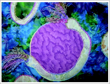
Figure 1. Quiltophagy.
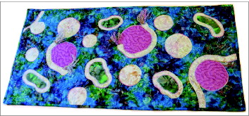
Figure 2. The quilter, Barbara M. Crumrine, displaying the final quilt as a wall hanging.
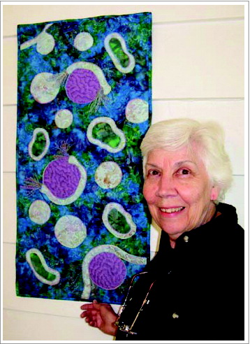
Figure 3. Background fabric depicting the cytosol, shown here with single- and double-membrane vesicles on top.
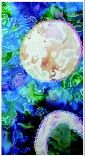
Figure 4. Assembly of a test pattern. (A) The artist in her sewing room assembling the pattern; yarn is shown on the bottom left of the sewing table. (B) The assembled test pattern.
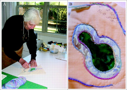
Figure 5. An intermediate stage showing (A) assembly of the autophagic components, and (B) a higher magnification demonstrating the couched stitch surrounding the membranes.
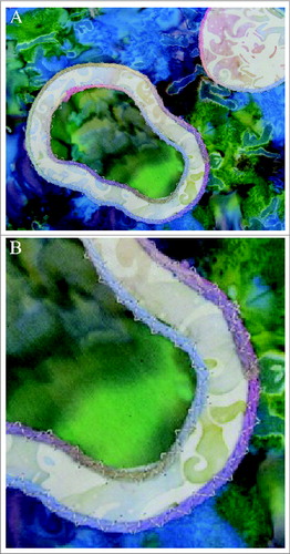
Figure 6. Detail depicting the growing ends of the phagophore.