Figures & data
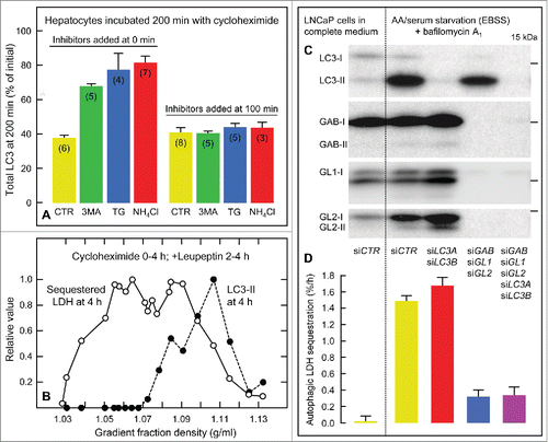
Figure 1. Experimental evidence for LC3-independent autophagy of cytosolic bulk cargo during starvation. (A) Turnover of LC3 in rat hepatocytes incubated for 200 min at 37°C with cycloheximide (100 μM) alone (CTR) or with 3-methyladenine (3MA, 10 mM), thapsigargin (TG, 5 μM) or NH4Cl (20 mM) added at 0 min (causing inhibition of degradative autophagic-lysosomal LC3 flux) or at 100 min (with no effect, indicating the absence of autophagic-lysosomal LC3 flux). Total LC3 remaining at 200 min is expressed as percent of the 0-min value (mean ±SE of the number of experiments given in parentheses). (B) Density gradient distribution of sedimentable hepatocytic organelles carrying autophagically sequestered LDH (open circles) or organelle-associated LC3-II (filled circles). The cells were harvested after 4 h of cycloheximide treatment, leupeptin (0.3 mM) having been added at 2 h to allow LDH accumulation during the period 2–4 h. (C) Effect of 52-h siRNA-mediated knockdown of the Atg8 homologs LC3A, LC3B, GABARAP (GAB), GABARAPL1 (GL1) and GABARAPL2 (GL2) on expression of the corresponding immunoblotted protein in LNCaP prostate cancer cells (siCTR, nontargeting siRNA; I, nonlipidated; II, lipidated form). The cells were incubated for 4 h in a complete medium or under amino acid- and serum-starved conditions (EBSS) in the presence of bafilomycin A1 (200 nM) as indicated. (D) Effects of the knockdowns on macroautophagic cargo (LDH) sequestration, expressed as percent of cellular LDH sequestered per h (mean ±SE of 3 experiments). Figures adapted from ref. Citation6.