Figures & data
Figure 1. Membrane scission and membrane fusion. (A) Membrane scission (or fission) is illustrated for the case where 2 equally sized bilayers are formed from one bilayer. Fusion is the topologically reverse process, where 2 bilayers or vesicles merge into a single one. (B, C) The size and location of the 2 vesicles relative to each other are not relevant to identify a process as scission or fusion. (D) Membrane scission and fusion of 2 almost equally sized vesicles, stacked within each other. The formation of the autophagosome from the cup-shaped phagophore provides an example for a scission process. Each bilayer is depicted as a single line, solid lines represent directly “visible” bilayers and dashed lines correspond to bilayers evident in cross sections only.
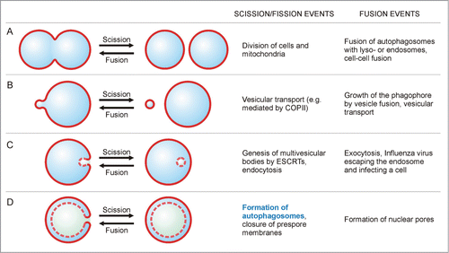
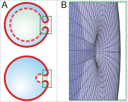
Figure 2. The scission neck. (A) The 2D cross-sectional views show 2 strongly bent membrane segments in the marked rectangular areas during autophagy (upper sketch) and endocytosis (lower sketch). (B) The rotated 3D view of the areas highlighted in (A) reveals that these 2 segments belong to a single scission neck.