Figures & data
Figure 1. Retinal degeneration in epg5 knockout mice. (A) Histological analysis of retinas from epg5−/− and Epg5+/− mice at the age of 3, 6, 10 and 15 mo. All sections were stained with hematoxylin-eosin and photographed from the first dorsal location (∼0.18 mm from the optic nerve head) at the same magnification (200 ×). Scale bars: 50 µm. RPE, retinal pigment epithelium; GCL, ganglion cell layer; IPL, inner plexiform layer; INL, inner nuclear layer; OPL, outer plexiform layer; ONL, outer nuclear layer; IS, inner segments; OS, outer segments. As a result of embedding, the RPE layer occasionally separates from the photoreceptor segments. (B) Temporal changes in retinal degeneration, presented as thickness of ONL, IS and OS normalized to INL thickness to correct for sectioning artifacts, in epg5−/− and Epg5+/− mice at the age of 3, 6, 10, 12 and 15 mo. Progressive decrease in the thickness of ONL, IS and OS is observed in epg5−/− retinas from 6 mo onwards. At 15 mo, the IS and OS are barely detected in epg5−/− mice. *P < 0.05, **P < 0.01, ***P < 0.001. Statistical comparisons were performed with 2-tailed, unpaired Student t tests. (C) The amplitudes of scotopic (dark-adapted) b-wave, mixed a-wave and mixed b-wave, and photopic (light-adapted) b-wave and flicker responses from epg5−/− and Epg5+/− mice at 6 mo (n = 6). *P < 0.05, **P < 0.01. Data were compared with 2-tailed, unpaired Student t tests.
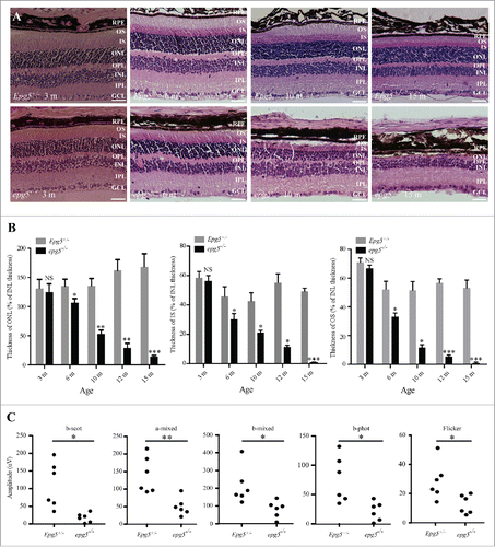
Figure 2. Accumulation of LC3-II, SQSTM1 aggregates and ubiquitin-positive inclusions in the retinas of epg5 knockout mice. (A) Immunoblot of LC3 and SQSTM1 in retinal extracts from epg5−/− and Epg5+/− mice aged 6 mo. (B) SQSTM1 aggregates dramatically accumulate in the GCL, OPL and INL of 3-mo-old epg5−/− mice, but not littermate controls. (C) Quantification of the number of cytoplasmic SQSTM1 and ubiquitin-positive aggregates in the retinas of epg5−/− and Epg5+/− mice aged 3 and 6 mo. Means ± SEM of 4 mice are shown. *P < 0.05, **P < 0.01. Statistical comparisons were performed with 2-tailed, unpaired Student t tests. (D) SQSTM1 aggregates and ubiquitin-positive inclusions dramatically accumulate and are colocalized in Epg5+/− retinas aged 6 mo. Scale bars in ((B)and D): 20 µm.
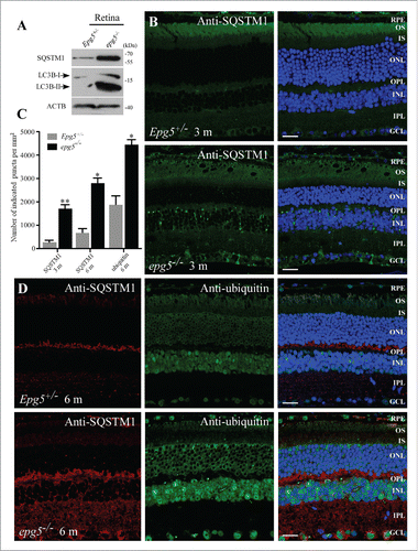
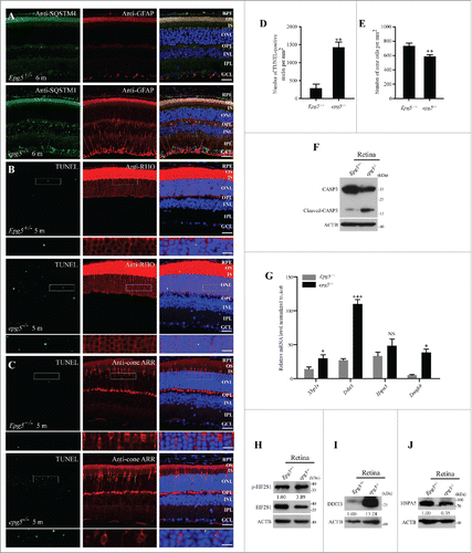
Figure 3. Photoreceptors undergo apoptosis and ER stress is activated in epg5−/− retinas. (A) Immunostaining analysis of cytoplasmic SQSTM1 and GFAP in the retinas of epg5−/− and Epg5+/− mice aged 6 mo. Few GFAP-stained structures are detected in Epg5+/− retinas, while many GFAP-stained tubular structures are observed throughout the retinas of epg5−/− mice. Scale bars: 20 µm. (B and C) TUNEL staining of 5-mo-old epg5−/− and Epg5+/− retinas that are costained with anti-RHO (rhodopsin) or anti-cone ARR/Arrestin antibodies. Scale bars: 20 µm. Enlargements of the boxed areas were shown on the bottom for each panel. Scale bars in enlargements: 60 µm. (D) Quantification of TUNEL-positive cells in retinas of epg5−/− and Epg5+/− mice. Means ± SEM of 6 paired mice at the age of 5 mo are shown. **P < 0.01. Statistical significance was determined by a 2-tailed, unpaired Student t test. (E) Quantification of anti-cone ARR-positive cones in epg5−/− and Epg5+/− mice at the age of 10 mo is shown in (E). Means ± SEM of 6 paired mice are shown. **P < 0.01. Statistical significance was determined by a 2-tailed, unpaired Student t test. Scale bars: 20 µm. (F) Immunoblotting analysis of full-length CASP3 and cleaved-CASP3 in retinas of epg5−/− and Epg5+/− mice at the age of 6 mo. Levels of cleaved CASP3 are dramatically increased in epg5−/− retinas compared with Epg5+/− retinas. (G) Transcriptional levels of genes involved in the UPR, including spliced Xbp1 (Xbp1s), Ddit3 and Eidj4, are significantly increased in epg5−/− retinas compared to controls at the age of 6 mo. *P < 0.05, ***P < 0.001. Data were compared with 2-tailed, unpaired Student t tests. (H, (I)and J) In epg5−/− retinas, DDIT3 and p-EIF2S1 protein levels are elevated, while the protein level of HSPA5, a downstream marker of the ATF6 pathway, is reduced.