Figures & data
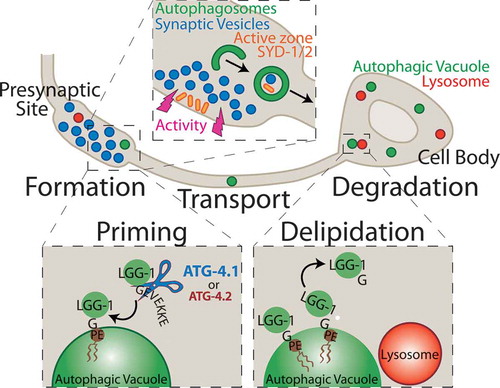
Figure 1. The schematic illustrates a model for synaptic autophagy across the cell biology of the neuron. Formation: Autophagosomes form at presynaptic sites dependent on neuronal activity, likely engulfing synaptic substrates including synaptic vesicle proteins and active zone proteins. Priming: Autophagy cysteine protease isoforms, primarily ATG-4.1, and also ATG-4.2, cleave the C-terminal end of LGG-1 to expose a terminal glycine residue, which is then conjugated onto the phagophore membrane at a phosphatidylethanolamine (PE) phospholipid to promote autophagosome formation. Transport: Autophagic vacuoles at the synapse are transported in a net retrograde fashion along the axon towards the cell body. Delipidation: The ATG-4.2 isoform, but not the ATG-4.1 isoform, cleaves LGG-1/2 off of the autophagosomal membrane to promote autophagosome maturation. Degradation: Autophagic vacuoles then fuse with acidic lysosomes and are degraded.